Monthly Archives: December 2024
CT Morphometric Analysis of Medial Femoral Condyle in Indian Population: An analytical study comparing Unicompartmental Knee Arthroplasty Implants to Indian knees.
Vol 10 | Issue 2 | July-December 2024 | page: 31-35 | Kedar Ahuja, Mohan Desai, Ibad Patel, Kannan Pughazendi, Ashraf Shaikh
https://doi.org/10.13107/jmt.2024.v10.i02.246
Author: Kedar Ahuja [1], Mohan Desai [1], Ibad Patel [1], Kannan Pughazendi [1], Ashraf Shaikh [1]
[1] Department of Orthopaedics, Seth GSMC and KEM Hospital, Mumbai, Maharashtra, India.
Address of Correspondence
Dr. Kedar Ahuja
Department of Orthopaedics, Seth GSMC and KEM Hospital, Mumbai, Maharashtra, India.
E-mail: drkedarahuja@gmail.com
Abstract
Background: The idea of resurfacing a single knee articular compartment was introduced in 1950s by MacIntosh and modified by McKeever who inserted a prosthetic disc into diseased tibial plateau, the disc functioned as a joint spacer effectively. Marmor designed the first unicompartmental knee prosthesis. However , early results were poor because of prosthetic loosening and osteoarthritis in the contralateral compartment. Osteoarthritis of the knee usually affects the medial compartment of the tibiofemoral articulation first and it may later involve the lateral compartment also [2,5]. Present study to compare dimensions of the medial femoral condyle with femoral components of conventional UKA prosthesis.
The purpose of this study was to compare dimensions of Medial Femoral Condyle (MFC) of Indian population to that of the standard size femoral components of commercially available three Unicompartmental Knee Arthroplasty (UKA) implants.
This is an analytical study wherein we included CT morphometric data of 100 consecutive nonarthritic adult knees with reference to the MFC, to assess the compatibility with UKA prostheses. A systematic approach was used to measure the anteroposterior dimension of each MFC. Proportion of knees which could be optimally replaced with the existing commercially available three Unicompartmental Knee Arthroplasty femoral implant systems viz., Link, DePuy and Smith & Nephew implants were calculated.
Results: There were 56 males and 44 females included in the study (M:F – 1.27:1). The mean age was 43.28 ± 10.53 years Best fit amongst the Link implant was the size ‘small’ in 43% of the patients, followed by ‘medium-small’ in 35% and ‘medium’ in 16%. For Depuy Sigma Uni implant, best fit was observed in size 1 in 38% of the patients, followed by size 3 in 25%, size 4 in 13%, size 2 in 10% and size 5 in 2%. Best fit among the Smith & Nephew implants was observed in the size 2 i.e, 30%, followed by size 3 in 27% , size 4 in 15%, size 1 in 12% and size 5 in 8%. A proportion of Indian Knees will have overhang of the femoral component of UKR because MFC of some Indian knees is smaller than the smallest size of the femoral component. which in our study was 6 % for Link, 12% for Depuy and 8% for Smith & Nephew.
Hypotheses: The best fit for Indian Knees is seen with the size implants ‘small’ in Link (43%), ‘size 1’ in Depuy implants (38%) and ‘size 2’ in Smith & Nephew implants (30%) of the patients. It is suggested that the implant companies should take the Indian population into consideration and customize the implants which may help in optimizing the fit and reducing overhang.
Clinical Importance: At If the femoral component is oversized it often causes anterior overstuffing that increases the risk of patellofemoral as well as soft-tissue impingement. So to encourage introduction of new ways to measure prosthesis sizes for Indian knee, or encourage companies to make sizes catering to Indian sizes.
Future Research: The shift towards personalized implant solutions could potentially revolutionize the field of knee arthroplasty, offering tailored interventions that better align with the diverse anatomical profiles of patients across the globe. Future directions in knee implant research should focus on large-scale, multi-center studies encompassing diverse populations
Conclusion: The best fit for Indian Knees is seen with the size implants ‘small’ in Link (43%), ‘size 1’ in Depuy implants (38%) and ‘size 2’ in Smith & Nephew implants (30%) of the patients.
Keywords: UKR, unicondylar knee replacement, partial knee replacement,medial compartmental osteoarthritis, Indian knees
Introduction:
Osteoarthritis is a common pathological disorder of the knee and mostly affects the medial compartment of the tibiofemoral articulation. [1,2] At present Unicompartmental Knee Arthroplasty (UKA) is the most beneficial treatment for unicompartmental knee arthritis because it is designed in a way that it preserves most of the patient’s own bone and thereby reducing trauma and improving post-operative recovery. [3] However If the femoral component is oversized it often causes anterior overstuffing that increases the risk of patellofemoral as well as soft-tissue impingement. Hence the successful outcome of UKA is mainly dependent on the best fit between the implant component and the resected surface of the knee and therefore it is imperative to keep in mind dimensional considerations before choosing the implants for UKA.
Almost all of the commercially available UKA implants are made as per the measurements available from the western population studies. [4] Currently there are no Indian studies available on the comparison of UKA implants to the components of the medial femoral condyle. And it is well known that Indians and westerners have predominantly striking racial differences with respect to various physical traits and dimensions.
The primary objective of this study was to compare the dimensions of medial femoral condyle i.e., Anteroposterior (AP) and Mediolateral (ML) diameters of Indian population with that of standard size femoral components of the commercially available UKA implants and secondary objective was to find out the most common sizes of the femoral implants that best fit the Indian Knees.
Methods:
This analytical, cross-sectional study was conducted at a tertiary care hospital following approval from the Institutional Ethics Committee (Project no. EC/223/2018). Sample size estimation and sampling technique was done as per Cohen’s d (effect size) calculation [5], assuming a significance level of 5% (α) and statistical power of 80% (1 − β), a sample size of 100 patients was calculated.
These patients underwent CT scan of their knee for various clinical indications excluding pathologies which could alter the morphometry of femoral condyle, i.e., fractures, neoplasia, old physeal injuries etc.
During the scan, it was ensured that the patients were in supine position, with the knees relaxed and in extension. The scanning procedure 3D- CT scan (120 kVp, 50 mAs, Philips Healthcare) was performed to acquire CT slices of thickness 0.5mm with a resolution of 512 × 512 pixels. The MFC dimensions were measured by a single surgeon using OsiriX DICOM (Digital Imaging and Communications in Medicine) viewer software for Mac OS.
Morphometric measurements of the Medial Femoral Condyle
A line was drawn along the posterior femoral cortex corresponding to the posterior peg of the UKA implant. The minimal resection is 6.7mm distally (distal femur), so a perpendicular line to posterior femoral cortex was drawn 6.7mm proximal to distal femoral cortex (Line A). Two more lines transecting the latter line at the anterior and posterior cortices are drawn parallel to each other at maximum AP (line 1 and 2). The distance between line 1 and line 2 was used to compare with different conventionally available implant sizes in AP. Mediolateral length was measured on the same axis (Line A) in axial cuts. Overhang was defined as Anteroposterior fit > 2 mm over and above the femoral condyle.
Statistical analysis
Patients’ demographic data, mediolateral and anteroposterior measurements of the MFC were summarized as mean and standard deviation (mean ± SD). The best-fit lines for each of the implant systems were calculated with the use of least squares regression method. Dimensions of the patients medial femoral condyle were then compared with various commercially available implants viz., Link , DePuy and the Smith & Nephew implants.
Statistical analysis was performed using Microsoft Excel and SPSS ver.20 (IBM Corp, Armonk, NY). Chi-square test of association was used to compare between the medial femoral condylar variables and the various types of UKA implants as well as to compare between males and females. The ‘p’ value ≤0.05 was considered to be statistically significant.
Results
There were 44 females and 56 males in this study, with a male: female ratio of 1.27: 1. Mean age of the study participants was 43.28 ± 10.53 years. Average ML diameter was 20.99 ± 1.44 and AP diameter was 47.56 ± 3.55. The minimum ML diameter was 17.3 and minimum AP diameter was 39.8.
The morphometric analysis revealed distinct trends in the best-fit sizes among the three commercially available Unicompartmental Knee Arthroplasty (UKA) implant systems: Link, DePuy, and Smith & Nephew.
Among the Link implant systems, the size 'small' emerged as the most suitable in 43% of the patients, demonstrating a notable prevalence for this category. Following closely, the 'medium-small' size exhibited an optimal fit in 35% of cases, emphasizing its relevance in a substantial proportion of the study cohort. The 'medium' size, although less common, still demonstrated a favorable fit in 16% of patients.
In the case of DePuy implants, size 1 took the lead as the best fit in 38% of patients, indicating its compatibility with a significant portion of the study population. Subsequent sizes demonstrated varying degrees of suitability, with size 3 showing an optimal fit in 25% of cases. Sizes 4, 2, and 5 exhibited successively diminishing percentages of optimal fits.
Among the Smith & Nephew implant systems, size 2 emerged as the most fitting choice in 30% of patients, closely followed by size 3 at 27%. The subsequent sizes—4, 1, and 5—showed descending percentages of optimal fits.
Despite the meticulous comparison with the smallest size implants available, a notable occurrence of femoral overhang was observed across all three implant systems. Specifically, 6 cases with Link implants, 12 with DePuy implants, and 8 with Smith & Nephew implants exhibited femoral overhang. This highlights a crucial aspect of the study, indicating that even with the smallest available implants, there remains a subset of patients for whom the femoral component extends beyond the resected surface of the knee. This emphasizes the imperative need for further refinement in implant design and customization to ensure an optimal fit for all patients, reducing the risk of complications associated with femoral overhang.
Discussion
Historical Development and Evolution of Unicompartmental Knee Arthroplasty (UKA):
The landscape of knee arthroplasty has undergone a transformative journey over the years, with significant advancements in implant designs and surgical techniques. Unicompartmental Knee Arthroplasty (UKA) emerged as a promising approach, offering a more conservative alternative to total knee replacement by preserving healthy tissues and promoting quicker postoperative recovery [3]. As osteoarthritis, particularly affecting the medial compartment of the knee, became a prevalent pathological disorder, the demand for effective and less invasive treatment options grew. The development of UKA was a response to this demand, aiming to provide a targeted solution for patients with unicompartmental knee arthritis.
The historical trajectory of UKA can be traced back to the pioneering work of John Insall and Chitranjan Ranawat in the 1970s, who introduced the concept of partial knee replacement to address isolated compartmental degeneration [3]. The Oxford meniscal knee, introduced by Carr et al. in 1993, marked a significant milestone in the evolution of UKA, demonstrating improved survival rates and functional outcomes [1]. Over the years, UKA has evolved from early fixed-bearing designs to modern mobile-bearing systems, enhancing implant stability and longevity. The evolution has also seen the introduction of various implant systems, each designed with specific features to optimize outcomes.
Challenges in Achieving Optimal Fit:
Despite the positive trajectory of UKA development, achieving an optimal fit between the implant components and the resected surface of the knee remains a complex task. Our study aligns with previous research, notably the work by Hitt et al [7], revealing the challenges in achieving comprehensive coverage of the exposed cortical rim with current knee arthroplasty systems. Notably, our findings indicate that even theoretically optimized implants fall short, covering only 76% of the cortical rim. This underscores the persistent complexity in achieving an ideal fit for knee implants and emphasizes the need for nuanced considerations in implant design.
Our investigation of three commercially available UKA implants sheds light on notable variations in overhang prevalence, with the lowest incidence observed in Link implants, followed by Smith & Nephew, and the highest in Depuy implants. The significant dominance of optimal fit in the Link Sled 'small' size (43%) highlights its close match to the medial femoral condyle of the Indian population, suggesting its potential superiority in achieving optimal outcomes [1].
Population-Specific Anatomical Variations:
The study emphasizes the critical importance of considering population-specific anatomical variations in implant design. The findings resonate with Cheng et al's examination of femoral implants in the Chinese population, which revealed overhang for all ranges of the anteroposterior (AP) dimension [8]. This parallel discovery underscores the importance of understanding population-specific anatomical variations to enhance implant design. Similarly, Fitz et al's study on cadavers emphasizes the disconnect between current UKA dimensions and measured sizes, particularly noting challenges in achieving an optimal fit, especially in males [6].
Gender-based anatomical differences, as explored by Yan et al [11], further highlight the intricacies of femoral morphology. The statistically significant gender-based variation in the ML/AP ratios of femoral condyles adds a layer of complexity to implant design considerations. In our study, the prevalence of overhang was generally higher in females across all three UKA implants, though it did not reach statistical significance.
Global Perspectives on Implant Design:
Studies on diverse populations, such as Kim et al's investigation into the femoral morphology of Korean women, emphasize the inadequacy of implant designs based on Caucasian population data [10]. Similarly, Surendran et al's research on the anthropometry of resected medial tibial condyles in Korean cadavers highlights challenges in achieving an optimal fit, with specific concerns regarding mediolateral overhang for certain tibial component designs [12]. The findings of Kantanavar et al's study on the compatibility of medial tibial condyle morphometry in the Indian population with contemporary UKA prostheses resonate with our results. [13].
Implications for Clinical Practice and Future Directions:
Our study contributes to the growing body of evidence emphasizing the need for population-specific considerations in knee implant design. The nuances of anatomical variations, as demonstrated by different studies, underscore the challenges in achieving a universally optimal fit. This collective evidence calls for a paradigm shift in implant design strategies, emphasizing customization to enhance the success and efficacy of Unicompartmental Knee Arthroplasty globally.
Furthermore, our study carries implications for clinical practice, suggesting that a one-size-fits-all approach to UKA implants may not be suitable, especially in diverse populations like India. The observed variations in optimal fit among different implants underscore the necessity for implant companies to consider the specific anatomical characteristics of the Indian population. Customizing implants based on regional anatomical variations may lead to improved patient outcomes, reduced complications, and enhanced long-term success of UKA.
This shift towards personalized implant solutions could potentially revolutionize the field of knee arthroplasty, offering tailored interventions that better align with the diverse anatomical profiles of patients across the globe. Future directions in knee implant research should focus on large-scale, multi-center studies encompassing diverse populations, including comprehensive data on anatomical variations and implant performance. Collaborations between researchers, surgeons, and implant manufacturers are essential to drive innovation, ensuring that the next generation of knee implants considers the unique characteristics of each patient population. In doing so, the field can move towards a more patient-centric approach, optimizing outcomes and quality of life for individuals undergoing Unicompartmental Knee Arthroplasty worldwide.
Conclusion
Our study results concluded that among the 3 commercially available implants compared, The best fit for Indian Knees is seen with the size implants ‘small’ in Link (43%), ‘size 1’ in Depuy implants (38%) and ‘size 2’ in Smith & Nephew implants (30%) of the patients. Through this study, we suggest a thorough consideration of population measurements in order to ensure best fit for majority of the Indian population. Since UKA is a long term treatment modality and prognosis is mainly dependent on how well the implants fit the patients morphology, we recommend a customized approach towards Indian population in planning of UKA components by commercial implant companies.
References
1. Carr A, Keyes G, Miller R, O’Connor J, Goodfellow J (1993) Medial Unicompartmental arthroplasty: a survival study of the Oxford meniscal knee. Clin Orthop 295:205–213
2. Lespasio MJ, Piuzzi NS, Husni ME, Muschler GF, Guarino A, Mont MA. Knee Osteoarthritis: A Primer. Perm J. 2017;21:16–183. doi: 10.7812/TPP/16-183.
3. Vasso M, Antoniadis A, Helmy N. Update on unicompartmental knee arthroplasty: current indications and failure modes. EFORT Open Rev. 2018;3(8):442–448. doi: 10.1302/2058-5241.3.170060.
4. Shah, D.S., et al. (2014) Morphological Measurements of Knee Joints in Indian Population: Comparison to Current Knee Prostheses. Open Journal of Rheumatology and Autoimmune Diseases, 4, 75-85
5. Fitz W, Bliss R, Losina E. Current fit of medial and lateral unicompartmental knee arthroplasty. Acta Orthop Belg. 2013 Apr;79(2):191-6. PMID: 23821971; PMCID: PMC4058334.//
6. Hitt K, Shurman JR II, Greene K, McCarthy J, Moskal J, Hoeman T, Mont MA (2003) Anthropometric measurements of the human knee: correlation to the sizing of current knee arthroplasty systems. J Bone Joint Surg Am 85:115–122
7. Cheng FB, Ji XF, Zheng WX, Lai Y, Cheng KL, Feng JC, Li YQ. Use of anthropometric data from the medial tibial and femoral condyles to design unicondylar knee prostheses in the Chinese population. Knee Surg Sports Traumatol Arthrosc. 2010 Mar;18(3):352-8. doi: 10.1007/s00167-009-0876-z. Epub 2009 Jul 24. PMID: 19629438.
8. Zhang Y, Wang X, Wu Z, Xia Q, Shao Y. Highly Variable Femoral Morphology in Osteoarthritic Chinese: Are Prostheses Today Sufficiently Suitable? J Knee Surg. 2017 Nov;30(9):936-942. doi: 10.1055/s-0037-1599250. Epub 2017 Mar 14. Erratum in: J Knee Surg. 2017 Nov;30(9):e1. PMID: 28293922.
9. Kim JB, Lyu SJ, Kang HW. Are Western knee designs dimensionally correct for Korean women? A morphometric study of resected femoral surfaces during primary total knee arthroplasty. Clin Orthop Surg, 2016, 8: 254–261.
10. Yan M., Wang J., Wang Y., Zhang J., Yue B., Zeng Y. Gender-based differences in the dimensions of the femoral trochlea and condyles in the Chinese population: correlation to the risk of femoral component overhang. Knee. 2014;21(1):252.
11. Mahoney O.M., Kinsey T. Overhang of the femoral component in total knee arthroplasty: risk factors and clinical consequences. J Bone Joint Surg Am. 2010;92(5):1115.
12. Surendran S, Kwak DS, Lee UY, Park SE, Gopinathan P, Han SH, Han CW. Anthropometry of the medial tibial condyle to design the tibial component for unicondylar knee arthroplasty for the Korean population. Knee Surg Sports Traumatol Arthrosc. 2007 Apr;15(4):436-42. doi: 10.1007/s00167-006-0188-5. Epub 2006 Sep 9. PMID: 16964513.
13. Kantanavar R, Desai MM, Pandit H. CT Morphometric Analysis of Medial Tibial Condyles: Are the Currently Available Designs of Unicompartmental Knee Arthroplasty Suitable for Indian Knees? Indian J Orthop. 2021 May 27;55(5):1135-1143. doi: 10.1007/s43465-021-00429-y. PMID: 34824713; PMCID: PMC8586401.
| How to Cite this Article: Ahuja K, Desai M, Patel I, Pughazendi K, Shaikh A. CT Morphometric Analysis of Medial Femoral Condyle in Indian Population: An analytical study comparing Unicompartmental Knee Arthroplasty Implants to Indian knees. Journal Medical Thesis. 2024 July-December; 10(2): 31-35. |
Institute Where Research was Conducted: Seth GSMC and KEM Hospital, Mumbai, Maharashtra,
India.
University Affiliation: Maharashtra University Of Health Sciences (MUHS), Nashik,
Maharashtra, India
Year of Acceptance of Thesis: 2020
Full Text PDF | Full Text HTML

A Study of Management of Grade IIIB Compound Fractures of Tibia/Fibula by Pirmary Ilizarov External Ring Fixator
Vol 10 | Issue 2 | July-December 2024 | page: 24-30 | Rajesh Gayakwad, Suhas S Shah
https://doi.org/10.13107/jmt.2024.v10.i02.244
Author: Rajesh Gayakwad [1], Suhas S Shah [1]
[1] Department of Orthopaedics, KB Bhabha Municipal General Hospital, Mumbai, Maharashtra, India.
[2] Department of Orthopaedics, GMC Healthcare, Mumbai, Maharashtra, India.
Address of Correspondence
Dr. Rajesh Gayakwad,
Consultant Orthopedic Surgeon,GMC Healthcare, Mumbai, Maharashtra, India.
Ex PG Resident, Department of Orthopaedics, KB Bhabha Municipal General Hospital, Mumbai, Maharashtra, India.
Email: drrajeshgaekwad@gmail.com
Abstract
Introduction: The current rationale for the management of open fractures is based on the work of Gustilo, Mendoza and Williams (1984). Their classification of such fractures has been shown to be of prognostic value (Caudle and Stern 1987; Hansen 1987). The prolonged nature of the treatment for type IIIB fracture frequently results in disruption of all aspects of the patient's life for a prolonged period.
Materials & Methods: There were 25 patients, with mean age of 29 years. The injuries were caused by motor vehicle accidents. All had type IIIB compound fractures of tibia/fibula. Patients received this treatment average 10 day post trauma. Management protocol included debridement, excision of devascularised bone ends, acute docking, bone grafting, and Ilizarov fixator with corticotomy. Patients were allowed to weight bear on 5th postoperative day. Average period of follow up was 2.5 yrs.
Results: All but three united, average time to union was 22 weeks. Average accepted LLD was 3cm (3 patients). 7 required second procedure in form of debridement and recorticotomy. There were 18 excellent, 3 good, 2 fair, 2 poor results. Complications encountered in form of pin tract infection (18% of pins), wire breakage (7), premature consolidation (1), delayed consolidation (2) osteomyelitis (6).
Discussion: We recommend the usage of Ilizarov apparatus to provide primary definitive fixation for high-energy long bone fractures. Early weight bearing even in severely comminuted fractures is the key factor that separates it from other methods of fixation. It promotes early functional recovery, eliminating fracture disease. Dynamisation and correction of deformities in any plane is easily accomplished. Frame constructs could be modified to facilitate wound cover and access. Therefore it lends the much-needed flexibility in complex fractures. Corticotomy, acute docking and lengthening allows removing the dead necrotic bone ends and correction of LLD by distraction at corticotomy site. In current study an association between the high energy trauma due to road traffic accident and incidence of Grade IIIb compound fractures was seen.
Conclusion: It is a one step solution, as it addresses simultaneously soft tissue cover to exposed bone, infection, and bone loss and permits early rehabilitation, thus good functional outcome.
Keywords: Compound tibia fractures, Wound Debridement, External Ring Fixator, Distraction osteogenesis, Corticotomy, Gustillo-Anderson score, AO Classification, DCP-Dynamic Compression Plate
Introduction:
Open fractures are more frequent in the tibia than any other long bone because one third of its surface is subcutaneous through most of its length. Furthermore the blood supply to the tibia is more precarious than that of other bones enclosed by heavy muscles. Due to all these factors, compound fractures of tibia are commonly complicated with delayed union, non-union and infection. More ever the optimal treatment of the compound fracture of shaft tibia remains controversial [1,2].
Thus treatment of the tibial shaft fractures has evolved from the era of life preservation to era of limb preservation and infection avoidance to era of optimum functional preservation. The appliances and methods available to treat the fractures of shaft tibia show the complexity and confusion in decision making with respect to the best treatment options.
In pre-listerian era, the compound fractures of tibia were mostly addressed by leg amputation in attempt to preserve life. With his principles of antisepsis, Joseph Lister managed these open injures with some success [3]. This heralded the approach of limb preservation and infection avoidance.
Throughout the 80’s external fixator had been the treatment of choice in open fractures of tibia as they provided stabilization with adequate access for wound management and soft tissue care. They had their own drawbacks in the form of pin track infection, pin loosening, malunion, delayed union and non-union. Plating as form of treatment devised by AO group is associated with increased risk of infection and skin necrosis [4,5].
Literature suggests that unreamed nails are superior to external fixator or the reamed nail in the management of compound fracture of tibia, while others did not find any difference between reamed and unreamed intramedullary nailing. Yet controversy exists on either side and rate of non-union with these procedures is too high near to about 37% [6,7,8]
Over the years Illizarov External Ring Fixation technique has been considered the ultimate solution for the compound fracture of tibia, both as primary modality and in those cases where the simple external fixators, reamed/unreamed intramedullary nailing methods have failed and in cases of non-union/malunion, considering the stability of the ilizarov apparatus which can be used to mobilize the patient at the earliest even in the severely comminuted fractures [9]
Hence this study endeavors to study the surgical technique of ilizarov external ring fixator and to evaluate clinical, functional and radiological results in the management of compound fractures of tibia caused by high energy trauma.
Materials And Methods
A total of 25 cases of Grade IIIb compound fractures of tibial/fibula shaft with fractures at least 7.5 cms from proximal articular surface and 5 cms from distal articular surface of tibia were selected. Patients with grade IIIc compound fractures, patients below 18 years and above 65 years, and those patients who found the apparatus aesthetically unacceptable were excluded from the study.
The Soft tissue component of the open wounds were classified with the one described by GUSTILLO-ANDERSON and modified by Gustillo, Gruninger and Davis [10,11], while the skeletal Component for the fractures of shat tibia-fibula, an AO Classification method was used [12].
After identifying the life threatening conditions and stabilizing the patient on the principles of ATLS[13], the wound in the injured limb was managed by removal of superficial contaminants, wound was washed with hydrogen peroxide, betadine so as to flush out blood clots, grit, contaminants .The wound was closed with sterile dressing soaked in normal saline. The deep exploration was done only in operation theatre.
Bactericidal intravenous antibiotic were started as soon as possible generally a third generation cephalosporin for gram positive coverage, an aminoglycoside for gram negative coverage, metronidazole for anaerobic coverage. Active and passive immunization against tetanus and clostridial organisms was given immediately. (Inj. ATS 1500 IV, Inj.AGGS 20,000 IV for compound fracture were administered as and when indicated) [14,15].
WOUDN IRRIGATION AND DEBIRDEMENT was done in the operation theatre under suitable anesthesia where thorough wound wash given with normal saline, hydrogen peroxide, betadine solution through mechanical irrigators like pulsatile lavage. The procedure was carried out under control of tourniquet with intermittent inflation and deflation to recognize the viability of the muscle and other soft tissues in the wound for which the quartet of Gregory was used, which includes-COLOR, CONSISTENCY, CAPACITY TO BLEED & CONTRACTILITY [16,17,18].
Definite treatment was attempted as early as possible within 24 hrs. In 19 patients the frame was applied within 6 hrs. Of another 6 patients, 4 patients presented to hospital with Monolateral ex-fix applied somewhere else. In other 2 patients, associated injuries did not allow immediate frame application. In them above knee slab was given with regular wound care till definitive surgery. The limb segment was sized with the actual ring or template with at least 2-3 cms of clearance between the inner edge of the ring and skin to accommodate the changes in dimensions of the extremity due to the swelling and dependent edema. More clearance was given posteriorly especially in the head injured and poly-trauma patients who were forced to be bedridden for a longer period of time. In our study, generally a 1600 ring size has met above criteria. The number of rings used was decided on the fracture anatomy and the dimensions of the open wound. Generally two rings above and two rings below the fracture site were used. Additional rings were applied as required which was decided on table. Six ring construct was done in segmental fractures (n-4, 16%).In rest of cases four ring construct was used. Bone grafting was done in patients with segmental fractures (n-4, 16%), fractures with bone loss (n-5, 20%) & rest of cases to augment healing (n-6, 24%). In extensively comminuted diaphyseal fractures, frame stability was enhanced by drop wires/shanz pins [19].
During surgery our aim was to make fracture site as transverse as possible. Special attention was given to the bleeding from the cortex of the fracture site and in cases of doubt, docking was done. Portable color Doppler was used to confirm the distal vascular status after acute docking in all cases of our study. After acute docking, the fracture fragments were held reduced by temporary four holed 4.5 mm DCP plate and cortical screws.
In our study, pure Russian technique was employed for frame construction [19]. A four-ring construct was generally applied in single level fractures with general rule of two level fixations in the major fracture fragment, generally we used a six ring construct. The distance between the fracture line and ring was kept to an average of 3 cms so as to increase the stability of the frame construct and to give enough room for compression/distraction and correction of any mal-alignment.
All wires used in our study were of olive type to add the stability of the construct and aid in fracture reduction and correction of any mal-alignment. Initially wires were passed in the shortest bone fragment parallel to the nearest joint to that short bone fragment. In mid shaft fractures, we preferred passing initial wires parallel to knee joint. Other wire is passed parallel to the opposite joint. These wires were used as reference wires for other wires. The angle formed between two wires passed at any level, was attempted to be at 90 degrees within the limits of the limb anatomy at that level. While transfixing the wires, power drill with start-stop technique with constant irrigation with cold saline by an assistant was used to avoid excessive thermal necrosis of the bone. Once the wire crosses the far cortex, drilling was stopped and wire was further pushed through by hammering with mallet or pulling out with pliers. During trans-fixation of wires, the muscles were kept at their maximum functional length and the nearby joints positions were changed when the wires passed from the flexor compartment to extensor compartment.i.e foot was dorsiflexed when anterior compartment was transfixed,platarflexed when posterior compartment was transfixed and inverted when peroneal compartment was impaled. The tension at skin-wire interface was kept to minimum either by mobilizing the skin in appropriate direction or by incising the skin at the interface. This was necessary to avoid any hindrance in joint range of motion [19].
Since we had used all of transfixing wire of olive type, tensioning was done from the side opposite to the olive bead. We used tension of 130 kg in all of the cases which was sufficient enough for weight bearing in our patients [20,21]. Following the surgical application of the frame, the various segments were moved by appropriate motors until, ideally, all rings were parallel and superimposed axially, and restoring the mechanical axis.
In our study corticotomy was done in case with extensive bone loss (n-5, 20%) during trauma or extesive bone removal(n-13, 52%) necessiated during process of docking.The level of corticotomy was at proximal metaphysis at level of tibial tuberosity between the two proximal full rings.We preformed the classical corticotomy used for triangular bones like tibia [22,23,24,25,26].
In our study, technique of wound healing by secondary intention and later closure by SSG was employed in most of patients (n-17, 68%). In very few patients (n-8, 32%) wound was managed by primary intention considering the severity of grade IIIb injuries. Of 8 cases, in only 2 patients wound could be closed by sutures [27,28]. In 5 cases wound was closed after docking at fracture site. In both instances wound was healthy enough for primary closure. In one case primary closure by local rotation flap was done with successful outcome. In 1(4%) patient local fascio-cutaneous flap was used to close the wound primarily since the wound status was good enough to allow the closure. This wound healed without infection. In another 6(24%) patients local rotation flaps of medial gastrocnemius (n-4, 16%) and soleus (n-2, 8%) was used with excellent result in form of complete healing of wound [29].
In patients with bone loss due to initial trauma, acute docking of fracture site, distraction at corticotomy site was started on POD-5 after check radiographs (n-18, 72%), at the speed of 1 mm/day which equals 1 complete rotation of the nut on the threaded connecting rod [30,31,32]. To aid this, the initial positions of the nut to be rotated were marked with paint (Nail polish) relative to their rings.
In all patients (n-25,100%) active /passive knee and ankle range of motion exercises are started on POD-1 onwards under supervision of physiotherapist. The physiotherapy session was done by the patient for about 2-3 hrs a day. On POD5, 4 patients(16%) with segmental fractures were kept NWB,while 7 patients (28%) kept PWB because of docking , >5 cms(n-3,12%) & bone loss with shortening <5 cms(4,16%).They proceeded to FWB by POD10.2 patient were still NWB till POD10 due bone loss >5 cms(1, 4%) and associated systemic injuries(1,4%) .These proceeded to PWB on POD5 and FWB on POD10.Majority of patients (21, 84%) were FWB by POD10 [33,34,35].
Results
23/25 fractures united. Average time to union was 22 weeks. Average accepted LLD was 3 cms in 3 patients.8/25 required second procedures. Second debridement was done in 3 cases of infected compound wounds closed primararily with sutures [36]. 1 patient had a Varus angulation of 5 degree at the fracture site (required correction of malalignment by recorticotomy and frame adjustment) [37]. 1 patient developed Stiffness around the knee joint; however the Knee range of motion was satisfactory in almost all cases. 3 cases of EHL transfixation occurred in our series, which later recovered after frame removal. In 2 cases wires were retensioned. In 2 cases recorticotomy was done for premature consolidation.1 patient required correction of loss of reduction on POD5 due to FWB.Second stage bone grafting was done in 5(20%) patients for delayed union, aseptic non-union (n-1,4%) and septic non-union(n-2,8%). Pin tract infection was seen in 7 (31) pins. All were grade I type which resolved by daily dressing and 5 day oral antibiotic course.1 case of foot drop was noticed in this series due to common peroneal nerve palsy during insertion of wire near knee joint. Wire passed posterior to neck of fibula. Spontaneous recovery occurred over 4 weeks without much active intervention. No vascular complications were seen any of the patients.
This study shows high incidence of open injuries in male population in comparison to female population, suggesting a relationship between the incidence and activity of the affected population. Our findings were comparable to standard epidemiological study [38,39,40,41] Middle third (n-14, 56%) followed by distal third (n-8, 32%) was most common anatomical locations of tibia affected in this study. Right leg was most commonly affected in our series. (n=18, 72%). There was clustering of the fractures in complex category (n-13, 52%, 42-c2.3).
In our study, 21(84%) cases sustained injury secondary to road traffic accidents. Majority of the victims were two wheeler riders followed by pedestrians. Only 1 case was of railway accident and 4 patients were of fall from height. The observation suggest that RTA is major causative agent of grade IIIb open injury with p>1, with the two wheeler riders and pedestrians being more susceptible to such injury. These findings are comparable to the findings of other studies. [38,39,40,41]
Based on Johner and Wruhs criteria [42], the final results were rated as 18 excellent, 3 good and 2 fair & 2 poor results were seen.
Discussion
In this study, Ilizarov fixator was chosen to primarily fix those fractures that produced high rate of complications with most conventional methods of fixation. For over half a century the Ilizarov device has been used for treatment of acute fractures and non-unions. This series is unique with respect to the complexity of the fractures considered. The specific category of fractures subjected to Ilizarov fixation was with the view to clearly define the role of the fixator in primary management of tibial fractures [43]. Even though the circumstances were adverse with respect to the fracture pattern, the union time was not unduly prolonged. Ilizarov external fixator provided one step solution in the management of compound fractures of long bones.
The structure is stable and enables the patient to bear weight on the affected limb even in much comminuted fractures, not easily achievable by other methods of fixation. The procedure is minimally invasive with little interference in the biology of fracture [44]. Current debate concerns the use of reamed and unreamed intramedullary nailing in open fractures because both techniques, to varying degrees negatively affect the circulation of cortical bone [45,46]. In patients with an open fracture, this has significant implications. Many standard studies have concluded against internal fixation of Grade III open fractures when compared to results of such fixation in Grade I, II open fractures. [46,47,48,49]. Based on literature, unreamed intramedullary nailing has been associated with a significantly lower rate of malunion than the external fixator or reamed nailing [50,51,52,53,54,55,56,57], nevertheless, unreamed nailing does not provide adequate stability for severely comminuted fractures with malunion rate of 0–27%.
Though IEF provides opportunity for simultaneous wound care and fracture management, it’s not as superior to monolateral external fixator because of technicality and cumbersome nature of ilizarov frame. Today the external fixator is considered only as temporary mode of fixation in compound fractures, which was done in 7 patients in our studies who had extensive soft tissue loss and contamination where primary IEF could not applied due to the technical aspects of IEF
Ilizarov is generally credited for the introduction of word corticotomy for percutaneous cortical osteotomy performed with osteotome. The word denotes an osteotomy of the cortex without disturbing the medullary canal and periosteum. It’s performed at metaphyseal level which according to ilizarov favor bone regenerate formation and also the soft tissues around this region are teleologically better adapted to distraction [58]. Monticello and Spinelli [59] and other orthopedic surgeons hold that the metaphysis has a much greater osteogenic potential than the diaphysis. Steen and Field [60] examined the difference between the healing of metaphyseal and diaphyseal level corticotomies under distraction. No difference was found in the healing time however the metaphyseal corticotomy was more stable than diaphyseal corticotomy [59,60].
However cases that required corticotomy and distraction, necessitated prolongation of the time on the fixator. However, this is an additional option with the Ilizarov fixator that makes it so versatile, since it allows correction of malalignment in three dimensions simultaneously also corrects LLD by principles of bone transport [61].
The elasticity of the wires used in the apparatus allows compressive movements at fracture site favoring rapid consolidation of the fracture callus, hence this method of treatment has shown high union rates in such complex fractures of bone which is known for non-union especially under influence of wound and high energy trauma [62,63,64]. Though axial compression can be achieved in intramedullary locked nails and monolateral external fixators, this is not possible in this class of fractures where high energy trauma causes comminution at fracture site. Schatzker reported 32 open tibial fractures treated with Ilizarov fixator. Healing time was 21.9 weeks in patients with a single injury and 25.7 weeks with multiple traumas similar to the results reported by Schwartzman et al. [65]. These features make this method of treatment unique when compared to other methods of treatment of open tibial fractures.
Conclusion
We recommend the usage of Ilizarov apparatus to provide primary definitive fixation for high-energy long bone fractures. Early weight bearing even in severely comminuted fractures is the key factor that separates it from other methods of fixation. It promotes early functional recovery, eliminating fracture disease. Dynamisation and correction of deformities in any plane is easily accomplished. Frame constructs could be modified to facilitate wound cover and access. Therefore it lends the much-needed flexibility in complex fractures. Corticotomy, acute docking and lengthening allows removing the dead necrotic bone ends and correction of LLD by distraction at corticotomy site. In current study an association between the high energy trauma due to road traffic accident and incidence of Grade IIIb compound fractures was seen.
References
1) Olson SA. Open fractures of the tibial shaft. In: Springfield DS, ed.Instructional Course Lectures Volume 46. Rosemont: AAOS; 1997:293-302.
2) Chapman MW, Olson SA: Open fractures. In: Rockwood CA Jr.,Green DP, Bucholz RW, et al. eds. Fractures in Adults, 4th ed.Philadelphia: Lippincott-Raven; 1996: 305-352.
3) Lister Joseph, 1867, A new method of treating compound fractures, abscesses, etc with observation on suppuration, LANCET 1, 326.
4) Aronson J. Factors influencing the choice of external skeletal fixation during distraction osteosynthesis. In Barr JJ, ed. Intructional course lectures, American academy of orthopaedic surgeons, St.Louis, MO, MOSBY; 1989; 38:175-183.
5) Alonso J, Geissler W, Hughes JL. External fixation of femoral fractures. Indications and limitations. Clin Orthop Relat Res 1989; 241:83-11). Murphy CP, D’Ambrosia RD, Dabezies EJ, Acker JH, Shoji H, Chuinard R.
6) Helfet DL, Howey T, Dipasquale T, et al. The treatment of open and/or unstable tibia fractures with an unreamed double locked tibial nail. Orthop Rev 1994; 23(suppl):9-17.
7) Sanders R, Jersinovich I, Angel, et al. The treatment of open tibial shaft fractures using an interlocked intramedullary nail without reaming. J Orthop Trauma 1994; 8:504-510.
8) Singer RW, Kellam JF. Open tibial diaphyseal fractures: Results of unreamed locked intramedullary nailing. Clin Orthop 1995; 9:77-120.
9) Macoy MT,Chao EYS,kasman RA.Comprision of mechanical performance in four type extrernal fixators. Clin orthop 1983; 180:20-23 11. Osteosynthesis, Berlin, Springer-Verlag; 1992: 369-45.
10) Gustillo RB, Mendoza RM, Williams DN. Problems in the management of type III (severe) open fractures: a new classification of type III open fractures.J Trauma. 1984; 24:742-6.
11) Brumback RJ, Jones AL. Inter observer agreement in the classification of open fractures of the tibia. The result of a survey of two hundred and forty-five orthopaedic surgeons. J Bone Joint Surg Am. 1994; 76:1162-6.
12) The AO classification of long bone fractures: an early study of its use in clinical practice. M L Newey 1, D Ricketts, L Roberts. Injury 1993 May;24(5):309-12.
13) American College of Surgeons Committee on Trauma (2004) Advanced trauma life support program for doctors, 7th edn. American College of Surgeons, Chicago
14) Stanford et al 1957, Role of prophylactic antibiotics in contaminated wounds, quoted by Harry F J. in J.B.J.S., 56A, No.3, 1974 of 541.
15) Patzakis, M. J.; Harvey, J. P., Jr.: And Ivler, Daniel: The Role of Antibiotics in the Management of Open Fractures.J. Bone and Joint Surg., 56-A: 532-541, April 1974.
16) Hampton OP, 1995, Basic principles in management of open fractures; J.A.M.A; 159, 41, 419, J Trauma, 1968, No.8.
17) Gross Arthur (1972); the technique of jet lavage in the severe open injuries, A.J.S; 12A; 373.
18) Delong WG Jr, Born CT, Wei SY, Petrik ME, Ponzio R, Schwab CW (1999) Aggressive treatment of 119 open fracture wounds. J Trauma 46:1049-1054. (s)
19) Catagni M (1991) A.S.AM.I (Association for the Study and Application of Ilizarov's Method) Group: Fractures of the Leg (Tibia). In Bianchi Maiocchi A, Aronson J (eds). Operative Principles of Ilizarov. Baltimore, Williams and Wilkins 91-124. (s)
20) Aronson J, Barry H, Boyd CM, Cannon DJ, Lubansky HJ.mMechanical induction of osteogenesis: The importance of pin rigidity. J Pdiatr Orthop. 1988;8:396-401
21) Ilizarov GA. The tension stress effect on the genesis and growth of tissue: Part I the influence of stability of fixation and soft tissue preservation. cu« Orthop. 1989;238:249-281.
22) Closed directed longitudino-oblique or spinal osteoclasia of the long tubular bones (experimental study). G A Ilizarov, P F Pereslitskikh, A P Barabash. Ortop Travmatol Protez . 1978 Nov:(11):20-3.
23) Illzarov, G. A., Kuznetsova, A. B., Peschansky, V. S., Schudlo, M. M., Khanes, G. S., Migalkins, N. S."Blood vessels under various regimens of the extremity distraction." Anat Gistol Embriol (Russian) 86:5; 4955,1984.
24) Monticelli G, Spinelli R. Leg lengthening by dosed metaphyseal corticotomy. ItaJJ Orthop Trallma'o/. 1983;9:139-150.
25) Steen H, Field T. Lengthening osteotomy in the metaphysis and diaphysis. Cun Orlhop. 1989;247:297-305.
26) Shtin VP, Nikiteno ET. Basing the term of beginning of distraction in operative lengthening of the leg in experiment.Ortop Travmatol Protez. 1974;35:5,48-51.
27) Cierny G III, Byrd HS, Jones RE (1983) Primary versus delayed soft-tissue coverage for severe open tibial shaft fractures. Clin Orthop 178:54-63.
28) Shtarker H, David R, Stolero J, Grimberg B, Soundry M (1997) Treatment of open tibial fractures with primary suture and Ilizarov fixation. Clin Orthop 335:268-274. (s)
29) Tukiainen E, Asko-Seljavaara S (1993) Use of the Ilizarov technique after free microvascular muscle flap transplantation in massive trauma of the lower leg. Clin Orthop 297:129-134. (s)
30) White SH, Kenwright J. The timing of distraction of an osteotomy.} Bone}oinJ Surg. 1990;72B:356-361.
31) Ilizarov GA. The tension stress effect on the genesis and growth of tissue: Part I the influence of stability of fixation and soft tissue preservation. cu« Orthop. 1989;238:249-281.
32) Perren SM,Huggler A,Russenberger M,Straumann F,Muller ME,Allgower M.A method of measuring change in compression applied to a living bone Acta orthop scand.1969;125(suppl):7-16
33) Goodship, A. E., Kenwright, J. "The influence of induced micro motion upon the healing of experimental tibial fractures." J Bone Joint Surg 678; 650-5, 85.
34) Lanyon, L. E., Rubin, C. T. "Static versus dynamic loads as an influence on bone remodeling." J Biomechanics12:897-907, 1984.
35) Bagnoli, G., Penna, F., Landini, A., Confalonieri, N., Torri, G., Pietrogrande, V. "Experimental study on bone formation under straining forces: Clinical and radiologic features." II Policlinico (Italian) 91:4, 908-13, 1984.
36) Baker JT ML, Costa AS, Nepola JV, Marsh LJ, Rodkey WA (1992) Comparison of infection rates in contaminated tibial fractures stabilized with internal vs external skeletal fixation in rabbits. J Orthop Trauma 6:509 (2007) 127:617–623 623
37) Ilizarov, G. A., Deviatov, A. A. "Operative elongation of the leg with simultaneous correction of deformities." Orthop Travrnatol Protez 30:3, 32-7, 1969.
38) The epidemiology of open long bone fractures. Court-Brown CM, Rimmer S, Prakash U, McQueen MM. Royal Infirmary of Edinburgh, UK Injury. 1998 Sep;29(7):529-34
39) Epidemiology Of Open Tibila Fractures In A Teaching Hosptials E B Ibenusi, A.U. Ekere. Department of surgery, University of port Harcourt teaching hospital, port Harcourt.
40) Injuries, Violence and Disabilities: biennial report 2006-2007 (2008): Reported road traffic fatalities in India (2006)-Who health organization Library.
41) “Road Accidents in India 2006.”: Ministry of Shipping, Road Transport and H highways
42) Classification of tibial shaft fractures and correlation with results after rigid internal fixation, R Johner, O Wruhs Clin Orthop Relat Res . 1983 Sep:(178):7-25.
43) Tucker HL, Kendra JC, Kinnebrew TE (1992) Management of unstable open and closed fractures using the Ilizarov Method. Clin Orthop 280:125-135. (s)
44) Macoy MT,Chao EYS,kasman RA.Comprision of mechanical performance in four type extrernal fixators. Clin orthop 1983; 180:20-23 11. Osteosynthesis, Berlin, Springer-Verlag; 1992: 369-45.
45) Rhinelander FW (1998) Effects of medullary nailing on the normal blood supply of diaphyseal cortex. Clin Orthop 350:5-17.
46) Schemitsch EH, Kowalski MJ, Swiontkowski MF (1996) Soft tissue blood flow following reamed versus unreamed locked intramedullary nailing:A fractured sheep tibia model. Ann Plast Surg 3:70-75.
47) Shannon FJ, Mullett H, O’Rourke K (2002) Unreamed intramedullary nail versus external Wxation in grade III open tibial fractures.J Trauma 52:650–654
48) Baker JT ML, Costa AS, Nepola JV, Marsh LJ, Rodkey WA (1992) Comparison of infection rates in contaminated tibial fractures stabilized with internal vs external skeletal fixation in rabbits. J Orthop Trauma 6:509 (2007) 127:617–623 623
49) Templeman DC, Gulli B, Tsukayama DT, Gustilo RB (1998) Update on the management of open fractures of the tibial shaft. Clin Orthop 350:18–25
50) Stegemann P, Lorio M, Soriano R, Bone L (1995) Management protocol for unreamed interlocking tibial nails for open tibial fractures.J Orthop Trauma 9:117–120
51) Alberts KA, Loohagen G, Einarsdottir H (1999) Open tibial fractures faster union after unreamed nailing than external Wxation Injury 30:519–523
52) Tu YK, Lin CH, Su JI, Hsu DT, Chen RJ (1995) Unreamed interlocking nail versus external Wxator for open type III tibia fractures.J Trauma 39:361–367
53) Santoro V HM, Benirschke S et al (1991) Prospective comparison of unreamed interlocking IM nails vs half pin external fixation and open tibial fractures. J. Orthop Trauma 5:238
54) Finkemeier CG, Schmidt AH, Kyle RF, Templeman DC (2000)Varecka TF A prospective, randomized study of intramedullary nails inserted with and without reaming for the treatment of open and closed fractures of the tibial shaft. J Orthop Trauma 14:187–193
55) Tornetta P 3rd, Bergman M, Watnik N, Berkowitz G, Steuer J(1994) Treatment of grade-IIIb open tibial fractures. A prospective randomised comparison of external fixation and non-reamed locked nailing. J Bone Joint Surg Br 76:13–19
56) Koval KJ, Clapper MF, Brumback RJ, Ellison PS Jr, Poka A, Bathon GH, Burgess AR (1991) Complications of reamed intramedullary nailing of the tibia. J Orthop Trauma 5:184–189
57) Bhandari M, Guyatt GH, Tong D, Adili A, Shaughnessy SG (2000) Reamed versus nonreamed intramedullary nailing of lower extremity long bone fractures: a systematic overview and metaanalysis. J Orthop Trauma 14:2–9
58) Ilizarov, G. A., Palienko, L. A, Shrejner, A A. "The bone marrow hemopoietic function and its relationship with the activity of osteogenesis upon reparative regeneration under the conditions of crus elongation in dogs." Ontogenez (Russian) 15:2, 1984. pp. 146-52.
59) Monticelli G, Spinelli R. Leg lengthening by dosed metaphyseal corticotomy. ItaJJ Orthop Trallma'o/. 1983;9:139-150.
60) Steen H, Field T. Lengthening osteotomy in the metaphysis and diaphysis. Cun Orlhop. 1989;247:297-305.
61) Kojimoto H, Yasui N, Gore T, Matsuda S, Shimomura Y. Bone lengthening in rabbits by callus distraction joint Bone Joint Surg.1988;70B:543-549.
62) Wolf, J., White, A., Punjabi, M., Southwick, W. "Comparison of cyclic loading versus constant compression in the treatment of long bone fractures in rabbits." J Bone Joint Surg 63A:805.g10, 1981.
63) Churches, A. E., Howlett, C. R. "The response of mature cortical bone to controlled time varying loading." : Mechanical Properties of Bone, Cowan (ed.), Sc. ASME Publication, AMD, Vol. 45, pp. 69-80.
64) Ilizarov, G. A., Palienko, L. A, Shrejner, A A Bogomjagkov, V. S. "Dynamics of the number of colony- forming cells for fibroblasts in the bone marrowand its relationship with the activity of osteogenesis upon the limb elongation." Ontogenez (Russian) 14:6, 1983. pp, 617-23
65) Schwartsman V, Martin SN, Ronquist RA, Schwartsman R (1992) Tibial fractures: the Ilizarov alternative. Clin Orthop 278:207-216.
| How to Cite this Article: Gayakwad R, Shah SS. A Study of Management of Grade IIIb Compound Fractures of Tibia/Fibula by Pirmary Ilizarov External Ring Fixator. Journal Medical Thesis 2024 July-December ; 10(2): 24-30. |
Institute Where Research was Conducted: K B Bhabha Municipal General Hospital, Mumbai,
Maharashtra, India.
University Affiliation: National Board of Examinations, New Delhi.
Year of Acceptance of Thesis: 2010

Full Text PDF | Full Text HTML
Validation of a Novel Clinico-Radiological Scoring System to Decide on the Need for Fusion in cases of Lumbar Degenerative Spondylolisthesis
Vol 10 | Issue 2 | July-December 2024 | page: 20-23 | Shashank Omprakashji Jajoo, Shailesh Hadgaonkar, Ajay Kothari, Siddharth Aiyer, Pramod Bhilare, Parag Sancheti, Ashok Kumar Shyam Murari, Darshankumar Sonawane
https://doi.org/10.13107/jmt.2024.v10.i02.242
Author: Shashank Omprakashji Jajoo [1], Shailesh Hadgaonkar [1], Ajay Kothari [1], Siddharth Aiyer [1], Pramod Bhilare [1], Parag Sancheti [1], Ashok Kumar Shyam Murari [1], Darshankumar Sonawane [1]
[1] Department of Orthopaedics, Sancheti Institute for Orthopaedics and Rehabilitation, Pune - 411005, Maharashtra, India.
Address of Correspondence
Dr. Shashank Omprakashji Jajoo,
Department of Orthopaedics, Sancheti Institute for Orthopaedics and Rehabilitation, Pune - 411005, Maharashtra, India
E-mail: shankrocks139.sj@gmail.com
Abstract
Background: Degenerative spondylolisthesis is a common cause of lower back pain, creating challenges in determining the best treatment approach—either standalone decompression or fusion. The absence of a standardized scoring system complicates decision-making. This study intends to validate a clinico-radiological scoring system to guide treatment decisions and improve patient outcomes.
Material & Methods: A cohort of 112 patients with degenerative lumbar spondylolisthesis was evaluated using the new scoring system. Independent assessments by spine consultants, fellows, and residents determined whether patients required standalone decompression or fusion. Inter- and intra-observer variability was measured. Patients' recovery and functional outcomes were tracked using VAS score (for back pain & leg pain), Oswestry Disability Index (ODI) and SF-36 score.
Results: A total of 112 cases were divided into four groups: Group 1A (8.9%), 1B (71.4%), 2A (13.4%), and 2B (6.3%). Complications were minimal, and re-surgery rates were low. Significant improvements were observed in back pain, leg pain, and ODI scores, with no major differences in postoperative outcomes across groups.
Conclusions: The scoring system effectively guides surgical decision-making in degenerative spondylolisthesis, reducing unnecessary fusion and improving outcomes. Further research should explore its broader application.
Keywords: Degenerative spondylolisthesis, Standalone decompression, Fusion, Scoring system, Reliability study
Introduction:
Degenerative spondylolisthesis has become a prominent cause of lower back pain and disability, especially as the global population ages and adopts more sedentary lifestyles. The management of this condition presents significant challenges for both patients and healthcare providers. A critical decision in treatment involves choosing between non-surgical approaches such as physical therapy, medications, or lifestyle changes, and opting for surgical intervention.[1] This decision is influenced by clinical, radiological, and patient-specific factors. However, a widely accepted standardized scoring system to guide these decisions is lacking. Furthermore, there is ongoing debate regarding the most appropriate surgical approach, with some advocating for decompression alone[2–10] and others supporting decompression combined with spinal fusion[2,11–13].
Several classification systems, including the Meyerding classification[14], Wiltse classification[15], and Clinical and Radiographic Degenerative Spondylolisthesis Classification (CARDS)[16], have been introduced to assist in surgical decision-making for degenerative lumbar spondylolisthesis. While these systems offer insights into spinal instability and the severity of the condition, they often fail to account for the complexities of individual cases. A key issue is the tendency to treat degenerative spondylolisthesis as a homogenous condition, leading to potential overtreatment or undertreatment[7]. For instance, the widely used Meyerding classification is limited as degenerative spondylolisthesis slips rarely exceed grade I or 30%[14]. This study seeks to validate a new clinico-radiological scoring system proposed by Kulkarni et al. in 2020[7] aimed at addressing these limitations and offering a more comprehensive, patient-centered approach to surgical decision-making in degenerative lumbar spondylolisthesis.
Aims and Objectives:
Aim: Validation of a Novel Clinico-Radiological Scoring System to Decide on the Need for Fusion in cases of Lumbar Degenerative Spondylolisthesis
Objectives: 1) Calculate the score for all patients with degenerative lumbar spondylolisthesis using the new scoring system. 2)Analyze and assess the functional outcomes of surgically treated patients. 3)Study the reliability of variables used in the new clinico-radiological scoring system. 4)Compare interobserver and intraobserver reliability of the new scoring system.
Materials and Methods:
This prospective study was conducted at a tertiary care center between October 2022 and December 2024. After receiving institutional ethical and scientific committee approval, patients were selected based on specific inclusion and exclusion criteria. Thorough explanations of the study's nature were provided to patients and their relatives, and informed consent was obtained from all participants. The sample size comprised approximately 112 skeletally mature patients diagnosed with degenerative lumbar spondylolisthesis.
Eligibility Criteria
Inclusion criteria: 1) Skeletally mature patients diagnosed with lumbar degenerative spondylolisthesis. 2) Patients who failed conservative treatment. 3) Patients with spondylolisthesis at one or two levels. 4) Patients who provided written informed consent.
Exclusion criteria: 1) Patients under 18 years of age. 2) Patients diagnosed with spondylolisthesis subtypes other than degenerative (e.g., dysplastic, isthmic, traumatic). 3) Patients previously managed surgically.
A new clinico-radiological scoring system proposed by Kulkarni et al. in 2020[7] was applied to calculate scores for all patients. Patients scoring <5.5 were classified as stable and advised standalone decompression. Scores ≥5.5 indicated instability, requiring fusion surgery[7].
Patients were divided into two main groups:
Group 1: Operated according to the new scoring system (Group 1A: standalone decompression, Group 1B: decompression with fusion).
Group 2: Operated based on the surgeon's preference, contrary to the scoring system (Group 2A: decompression with fusion, Group 2B: standalone decompression).
Postoperative follow-ups were conducted at 6 weeks, 3 months, 6 months, and 1 year. Functional outcomes were measured using VAS, ODI, and SF-36 Health Survey scores. Intra-operative and post-operative complications were monitored.
Seven independent observers were selected to evaluate a set of clinical cases twice, at intervals of 2-3 months, for interobserver and intraobserver reliability. Observers were blinded to each other’s assessments and their prior evaluations. The data was analyzed using Cohen’s Kappa statistic[16] to assess both inter-observer and intra-observer reliability. Kappa (k) values, expressed with 95% confidence intervals, ranged from -1 to 1, with higher values indicating better agreement.
Statistical analysis was performed using SPSS version 24.0. Comparisons were conducted using the Chi-Square test for categorical data and ANOVA for continuous variables, with Bonferroni post-hoc tests for multiple comparisons[17–19].
Results:
A total of 112 cases were analyzed and categorized into four groups: Group 1A (10 cases), Group 1B (80 cases), Group 2A (15 cases), and Group 2B (7 cases). The majority of cases belonged to Group 1B (71.4%), followed by Group 2A (13.4%), Group 1A (8.9%), and Group 2B (6.3%). The highest mean age was observed in Group 2A (67.80 ± 8.15 years), with a significant age difference between Group 2A and Group 1B (P<0.05). The male-to-female ratio in the study was 0.75:1. Group 2B had a significantly higher proportion of male patients (85.7%), while Group 1B had the largest proportion of female patients (61.2%).
BMI varied across the groups, ranging from 24.91 ± 4.45 kg/m² in Group 1A to 26.15 ± 2.94 kg/m² in Group 2B, but no significant differences were found (P>0.05). Co-morbidities such as hypertension, diabetes, ischemic heart disease (IHD), and hypothyroidism were prevalent.
Intra-operative complications occurred in 8.8% of Group 1B cases and 20% of Group 2A cases, primarily dural tears. Post-operative complications, including infection and cage migration, were minimal, occurring in 6.2% of Group 1B and 6.7% of Group 2A cases. No complications were reported in Groups 1A and 2B. Re-surgery was required in 2.5% of Group 1B and 6.7% of Group 2A cases, while Groups 1A and 2B had no re-surgeries.
Back pain, leg pain, Oswestry Disability Index (ODI), and SF36 scores were analyzed to assess outcomes. Group 1A had significantly lower pre-operative back pain scores (Mean = 3.70, SD = 3.09) compared to other groups (P<0.05). However, post-operative scores showed no significant differences at 6 weeks, 3 months, 6 months, and 1 year (P>0.05). The percentage improvement in back pain scores at 1 year ranged from 64.95% in Group 1A to 78.11% in Group 1B, with no significant differences between groups (P>0.05).
For leg pain, Group 1A had significantly higher pre-operative scores (Mean = 8.60, SD = 0.84) than Group 1B (Mean = 6.76, SD = 1.59), with no significant differences between other groups. At the 1-year follow-up, leg pain scores were significantly lower in Groups 1B and 2B compared to Group 2A (P<0.05), with percentage improvement ranging from 64.64% in Group 2A to 88.76% in Group 2B.
ODI scores were similar across all groups pre-operatively. At the 1-year follow-up, Groups 1A and 1B had significantly better scores compared to Group 2A (P<0.05), with percentage improvement ranging from 46.52% in Group 2A to 54.50% in Group 1B.
SF36 pain scores showed no significant pre-operative differences between the groups (P>0.05). At 1 year, Group 2B had the greatest improvement, with a 398.89% increase in pain scores, followed by Group 1A (368.15%), Group 1B (330.83%), and Group 2A (264.81%). Physical functioning scores also improved significantly across all groups post-operatively, with Group 2B showing the greatest improvement at 1 year (257.14%).
The interobserver agreement for parameters such as Mechanical Back Pain, age, and activity showed very high reliability, with Cohen's kappa values ranging from 0.999 for MBP and activity to 0.687 for Arvind’s score. Segmental Kyphosis and Facet Effusion had substantial agreement (kappa values ranging from 0.630 to 0.946). However, variability was noted in the assessment of Segmental Dynamic Spondylolisthesis (kappa values of 0.379 to 0.682), and technical factors showed the lowest agreement (kappa range 0.323 to 0.718). Intraobserver reliability mirrored these trends, with high agreement across most parameters, though certain parameters like Arvind's score and technical factors displayed slight variability.
Conclusion:
The results of the study showed significant improvements in patients who underwent surgical treatment, whether it was standalone decompression or decompression with fusion. The study further compared these findings with Kulkarni's study[7], highlighting similar trends in the reduction of pain scores, though with varying degrees of improvement. The inter-observer reliability of the scoring systems used in this study is generally high, certain parameters, particularly those involving more subjective assessments, could benefit from further refinement to enhance consistency. The robust agreement in most parameters underscores the reliability of the scoring systems, yet highlights the importance of continuous evaluation and training to ensure the highest standard of clinical assessments. In conclusion, the study validates the efficacy of the new clinico-radiological scoring system, which could potentially standardize the decision-making process in surgical treatment of degenerative spondylolisthesis, ensuring better patient outcomes and minimizing unnecessary fusion surgeries.
Clinical Message:
The validation of a new clinico-radiological scoring system to determine the need for fusion holds significant clinical importance and have potential of transforming the management of degenerative spondylolisthesis. The scoring system standardizes the decision-making process, reducing the variability that currently exists among surgeons. This standardization ensures that patients receive consistent and appropriate care, regardless of the treating surgeon.
A subgroup of patients with Degenerative Spondylolisthesis can get away with just stand-alone decompression, without the need of fusion which is more morbid surgical intervention. This have benefits of reduced surgical risk, reduced surgical time, shorter recovery time, preservation of motion, lower cost of surgery, etc. By accurately identifying patients who can benefit from decompression alone, the system helps avoid unnecessary fusion surgeries, thereby minimizing the associated morbidity and healthcare expenses.
References
1. Weinstein JN, Lurie JD, Tosteson TD, Hanscom B, Tosteson ANA, Blood EA, et al. Surgical versus Nonsurgical Treatment for Lumbar Degenerative Spondylolisthesis. N Engl J Med. 2007 May 31;356(22):2257–70.
2. Chan AK, Bisson EF, Bydon M, Glassman SD, Foley KT, Potts EA, et al. Laminectomy alone versus fusion for grade 1 lumbar spondylolisthesis in 426 patients from the prospective Quality Outcomes Database. J Neurosurg Spine. 2019 Feb;30(2):234–41.
3. Försth P, Ólafsson G, Carlsson T, Frost A, Borgström F, Fritzell P, et al. A Randomized, Controlled Trial of Fusion Surgery for Lumbar Spinal Stenosis. N Engl J Med. 2016 Apr 14;374(15):1413–23.
4. Cheung JPY, Cheung PWH, Cheung KMC, Luk KDK. Decompression without Fusion for Low-Grade Degenerative Spondylolisthesis. Asian Spine J. 2016;10(1):75.
5. Inose H, Kato T, Yuasa M, Yamada T, Maehara H, Hirai T, et al. Comparison of Decompression, Decompression Plus Fusion, and Decompression Plus Stabilization for Degenerative Spondylolisthesis: A Prospective, Randomized Study. Clin Spine Surg Spine Publ. 2018 Aug;31(7):E347–52.
6. Dijkerman ML, Overdevest GM, Moojen WA, Vleggeert-Lankamp CLA. Decompression with or without concomitant fusion in lumbar stenosis due to degenerative spondylolisthesis: a systematic review. Eur Spine J. 2018 Jul;27(7):1629–43.
7. Kulkarni AG, Kunder TS, Dutta S. Degenerative Spondylolisthesis: When to Fuse and When Not to? A New Scoring System. Clin Spine Surg Spine Publ. 2020 Oct;33(8):E391–400.
8. Ha DH, Kim TK, Oh SK, Cho HG, Kim KR, Shim DM. Results of Decompression Alone in Patients with Lumbar Spinal Stenosis and Degenerative Spondylolisthesis: A Minimum 5-Year Follow-up. Clin Orthop Surg. 2020;12(2):187.
9. Austevoll IM, Hermansen E, Fagerland MW, Storheim K, Brox JI, Solberg T, et al. Decompression with or without Fusion in Degenerative Lumbar Spondylolisthesis. N Engl J Med. 2021 Aug 5;385(6):526–38.
10. Wei FL, Zhou CP, Gao QY, Du MR, Gao HR, Zhu KL, et al. Decompression alone or decompression and fusion in degenerative lumbar spondylolisthesis. eClinicalMedicine. 2022 Sep;51:101559.
11. Herkowitz HN, Kurz LT. Degenerative lumbar spondylolisthesis with spinal stenosis. A prospective study comparing decompression with decompression and intertransverse process arthrodesis. J Bone Joint Surg Am. 1991 Jul;73(6):802–8.
12. Kleinstueck FS, Fekete TF, Mannion AF, Grob D, Porchet F, Mutter U, et al. To fuse or not to fuse in lumbar degenerative spondylolisthesis: do baseline symptoms help provide the answer? Eur Spine J. 2012 Feb;21(2):268–75.
13. Ghogawala Z, Dziura J, Butler WE, Dai F, Terrin N, Magge SN, et al. Laminectomy plus Fusion versus Laminectomy Alone for Lumbar Spondylolisthesis. N Engl J Med. 2016 Apr 14;374(15):1424–34.
14. Meyerding HW. Meyerding HW. Spondylolisthesis. Surg Gynecol Obstet. 1932;54: 371–377. In p. 371–7.
15. Wiltse LL, Newman PH, Macnab I. Classification of spondylolisis and spondylolisthesis. Clin Orthop. 1976 Jun;(117):23–9.
16. McHugh ML. Interrater reliability: the kappa statistic. Biochem Medica. 2012;22(3):276–82.
17. Rosner BA. Fundamentals of biostatistics. 5. ed. Pacific Grove, Calif.: Duxbury; 2000. 80–240 p.
18. Riffenburgh RH. Statistics in medicine. 2nd ed. Amsterdam: Elsevier Academic Press; 2006. 85–125 p.
19. Sundar Rao PSS, Richard J. An introduction to biostatistics: a manual for students in health sciences. 3 ed., 8. print. New Delhi: Prentice Hall of India; 2003. 86–160 p. (Eastern economy edition).
| How to Cite this Article: Jajoo SO, Hadgaonkar S, Kothari A, Aiyer S, Bhilare P, Sancheti P, Murari AS, Sonawane D. Validation of a Novel Clinico-Radiological Scoring System to Decide on the Need for Fusion in cases of Lumbar Degenerative Spondylolisthesis. Journal of Medical Thesis. 2024 July-December ; 10(2): 20-23. |
Institute Where Research was Conducted: Department of Orthopaedics, Sancheti Institute for Orthopaedics and Rehabilitation, Pune, Maharashtra, India.
University Affiliation: Maharashtra University Of Health Sciences (MUHS), Nashik, Maharashtra, India.
Year of Acceptance of Thesis: 2024

Full Text PDF | Full Text HTML
Functional and Radiological Outcome Comparison for Plating Vs Nailing Fixation in Closed Extra-Articular Tibia Fractures
Vol 10 | Issue 2 | July-December 2024 | page: 17-19 | Shrikant Pradeep Savant
https://doi.org/10.13107/jmt.2024.v10.i02.240
Author: Shrikant Pradeep Savant [1]
[1] Department of Orthopaedics and Joint Replacement, Criticare Asia Hospital, Andheri East, Mumbai, Maharashtra , India
Address of Correspondence
Dr. Shrikant Pradeep Savant,
Consultant Orthopaedics and Joint Replacement, Criticare Asia Hospital, Andheri East, Mumbai, Maharashtra ,
India.
E-mail: shrikantsavant11@gmail.com
Abstract
Purpose: The treatment of tibia fractures remains a gray area due to its tendency to have different modalities for different fracture patterns, a high nonunion and malunion rate and its tendency for infection due to its proximity to the skin. We conducted a study to compare the results of tibia fractures in terms of the rate of union, quality of the limb, incidence of complications and patient satisfaction.
Methods: Patients with extra articular tibia fractures, who were admitted in a tertiary care hospital from July 2017 to May 2019 taken for study after obtaining their consent. Follow up of the cases done for a period of 24 months. Exclusion criteria included medically unfit patient for surgery, compound fractures and pathological fractures.
Conclusion: Fracture type or pattern does not influence the eventual functional outcome.Closed reduction with tibia interlocking nail has got lesser early as well as late post operative complications as compared internal fixation with plating. Closed reduction with interlocking nail has got the advantage of early mobilization with full weight bearing as compared to with plating in which weight bearing is delayed.
Keywords: Tibia shaft fracture, Extra articular, Nailing vs plating, LEFS score
Introduction
The tibia, commonly known as the shinbone, is the larger and stronger bone in the lower leg. It bears the full weight of the body and connects the knee joint to the ankle joint. Despite its strength, the tibia is the most commonly fractured long bone, due to its location, orientation, and lack of soft tissue protection. Tibial diaphyseal fractures are particularly common, with an incidence of approximately 26 per 100,000 people per year.
In recent times, closed reduction with intramedullary interlocking nailing has become the preferred method for managing tibial fractures in adults. This technique offers mechanical stability, better alignment, and preserves the biological environment of the fracture, allowing soft tissues to remain intact. Locking plate osteosynthesis is typically used for fractures that extend into the metaphysis, though complications like infection, wound breakdown, malunion, and nonunion are frequently observed.
With both techniques being widely used, the choice of treatment for tibial fractures remains a subject of debate. This study aims to compare the outcomes of these two methods in terms of fracture union, limb quality, complication rates, and patient satisfaction.
Aims and Objective
The primary objective of this study is to compare the clinical outcomes of two surgical methods used to treat tibial fractures: intramedullary interlocking nailing and locking plate osteosynthesis. The study aims to assess the following:
1. Union Rate: Compare the time required for fracture union between the two techniques.
2. Functional Outcome: Evaluate the functional outcome using the Lower Extremity Functional Scale (LEFS).
3. Complication Rate: Identify early and late post-operative complications for both treatment options.
4. Patient Satisfaction: Measure patient satisfaction at follow-ups using clinical and functional parameters.
Materials and Methods
Source of Data
This prospective study was conducted on 46 patients with extra-articular tibia fractures, admitted to a tertiary care hospital from July 2017 to May 2019. Patients were followed for 24 months across six visits (6 weeks, 3 months, 6 months, 12 months, 18 months, and 24 months). The study was conducted after obtaining ethical approval from the institutional review board and informed consent from all patients.
Inclusion Criteria
- Patients with closed extra-articular tibial fractures.
- Patients with a mature skeleton.
- Patients medically fit for surgery.
Exclusion Criteria
- Medically unfit patients.
- Compound fractures.
- Pathological fractures.
Protocol
Upon admission, a detailed examination and routine pre-operative investigations were performed. X-rays (AP and lateral views) of the tibia, knee, and ankle were taken. After obtaining pre-anaesthetic clearance, patients underwent either closed reduction with intramedullary interlocking nailing or open reduction and internal fixation with locking plate osteosynthesis. The choice of surgical technique was based on fracture type and surgeon preference.
Functional outcomes were assessed using the Lower Extremity Functional Scale (LEFS), with scores ranging from 0 to 80. Higher scores indicate better functional outcomes.
Statistical Analysis
Data was analyzed using SPSS-17. Chi-square and Fischer’s exact tests were used for qualitative data, while Student’s t-test was used for quantitative data. A p-value of <0.05 was considered statistically significant.
Results
1) Demographics: Among the 46 patients, 36 were males (78.26%) and 10 were females (21.74%). The most common age group was 51-60 years (28.26%).
2) Fracture Types: Proximal third tibial fractures were the most common (60.86%), followed by distal and mid-shaft fractures (19.57% each).
3) Mode of Injury: The majority of injuries were caused by road traffic accidents (65%), followed by railway accidents (35%).
4) Associated Injuries: Fibular fractures were the most common associated injury (59.45%).
5) Surgical Intervention: Tibial interlocking nailing was performed in 36.95% of cases, while tibial plating was performed in 45.65% of cases.
6) Complications
Early post-operative complications (e.g., swelling, bleb formation) were more common with plating (27.58%) compared to nailing (11.76%).
Late post-operative complications (e.g., wound dehiscence, infection) were significantly higher in the plating group compared to the nailing group.
7) Functional Outcome: The LEFS score was above 40 in 82.62% of patients, with better results observed in the nailing group.
8) Union Rates: Radiological union occurred within 90 days in 88.23% of nailing cases compared to 44.82% of plating cases. This difference was statistically significant (p=0.0093).
Conclusion
The study suggests that intramedullary interlocking nailing is a more favorable option for treating tibial fractures due to its lower complication rates, faster time to union, and better functional outcomes as measured by the LEFS. Plating, while effective for certain fracture types (e.g., fractures extending into the metaphysis), is associated with higher rates of early and late complications, such as wound infection and implant exposure.
Clinical Message
The results of this study have important implications for clinical practice. Intramedullary interlocking nailing should be the preferred treatment for extra-articular tibial fractures due to its superior outcomes in terms of union time, fewer complications, and better functional recovery. Surgeons should consider patient factors and fracture characteristics, but this study supports the widespread adoption of tibial nailing, especially in cases where early mobilization is desired.
References
1. He GC, Wang HS, Wang QF, Chen ZH, Cai XH. Effect of minimally invasive percutaneous plates versus interlocking intramedullary nailing in tibial shaft treatment for fractures in adults: a meta-analysis. Clinics. 2014;69(4):234-40.
2. Lindvall E, Sanders R, DiPasquale T, Herscovici D, Haidukewych G, Sagi C. Intramedullary nailing versus percutaneous locked plating of extra-articular proximal tibial fractures: comparison of 56 cases. Journal of orthopaedic trauma. 2009 Aug 1;23(7):485-92.
3. Vallier HA, Cureton BA, Patterson BM. Randomized, prospective comparison of plate versus intramedullary nail fixation for distal tibia shaft fractures. Journal of orthopaedic trauma. 2011 Dec 1;25(12):736-41.
4. Kwok CS, Crossman PT, Loizou CL. Plate versus nail for distal tibial fractures: a systematic review and meta-analysis. Journal of orthopaedic trauma. 2014 Sep 1;28(9):542-8.
5. Mao Z, Wang G, Zhang L, Zhang L, Chen S, Du H, Zhao Y, Tang P. Intramedullary nailing versus plating for distal tibia fractures without articular involvement: a meta-analysis. Journal of orthopaedic surgery and research. 2015 Dec;10(1):95.
6. Meena RC, Meena UK, Gupta GL, Gahlot N, Gaba S. Intramedullary nailing versus proximal plating in the management of closed extra-articular proximal tibial fracture: a randomized controlled trial. Journal of Orthopaedics and Traumatology. 2015 Sep;16(3):203.
7. Saied A, Ostovar M, Mousavi AA, Arabnejhad F. Comparison of intramedullary nail and plating in treatment of diaphyseal tibial fractures with intact fibulae: A randomized controlled trial. Indian journal of orthopaedics. 2016 May;50(3):277.
8. Mukherjee S, Arambam MS, WaikhOM S. Interlocking Nailing Versus Plating in Tibial Shaft Fractures in Adults: A Comparative Study. Journal of Clinical and Diagnostic Research: JCDR. 2017 Apr;11(4):RC08.
9. Daolagupu AK, Mudgal A, Agarwala V, Dutta KK. A comparative study of intramedullary interlocking nailing and minimally invasive plate osteosynthesis in extra articular distal tibial fractures. Indian journal of orthopaedics. 2017 May;51(3):292.
10. Mohindra M, Jain JK. Fundamentals of orthopedics. JP Medical Ltd; 2017 Dec 31.
11. Prasad PN, Nemade A, Anjum R, Joshi N. Extra-articular distal tibial fractures, is interlocking nailing an option? A prospective study of 147 cases. Chinese Journal of Traumatology. 2019 Apr 1;22(2):103-7.
12. Yao Q, Ni J, Peng LB, Yu DX, Yuan XM. Locked plating with minimally invasive percutaneous plate osteosynthesis versus intramedullary nailing of distal extra-articular tibial fracture: a retrospective study. Zhonghua yi xue za zhi. 2013 Dec;93(47):3748-
13. Huang P, Tang PF, Yao Q, Liang YT, Tao S, Zhang Q, Guo YZ, Liang XD, Wang Y. A comparisive study between intramedullary interlocking nail and plate-screw fixation in the treatment of tibial shaft fractures. Zhongguo gu shang= China journal of orthopaedics and traumatology. 2008 Apr;21(4):261-3.
14. Guo JJ, Tang N, Yang HL, Tang TS. A prospective, randomised trial comparing closed intramedullary nailing with percutaneous plating in the treatment of distal metaphyseal fractures of the tibia. The Journal of bone and joint surgery. British volume. 2010 Jul;92(7):984-8.
15. Gao WQ, Hu JH, Gu ZC, Zhang HX, Min P, Zhang LJ, Yu WW, Wang GL. Comparison of effect between early and delayed in primary intramedullary nailing combined with locked plate fixation for the treatment of multi-segments tibial fractures of type. Zhongguo gu shang= China journal of orthopaedics and traumatology. 2015 Feb;28(2):122-5.
16. Li Y, Jiang X, Guo Q, Zhu L, Ye T, Chen A. Treatment of distal tibial shaft fractures by three different surgical methods: a randomized, prospective study. International orthopaedics. 2014 Jun 1;38(6):1261-7.
17. Kuhn S, Appelmann P, Pairon P, Mehler D, Rommens PM. The Retrograde Tibial Nail: presentation and biomechanical evaluation of a new concept in the treatment of distal tibia fractures. Injury. 2014 Jan 1;45:S81-6.
18. Kiel J, Kaiser K. Stress reaction and fractures. InStatPearls [Internet] 2019 Jun 4. StatPearls Publishing.
19. Nelson Jr GE, Kelly PJ, Peterson LF, Janes JM. Blood supply of the human tibia. JBJS. 1960 Jun 1;42(4):625-36.
20. Jastifer JR, Gustafson PA, Labomascus A, Snoap T. Ball and socket ankle: mechanism and computational evidence of concept. The Journal of Foot and Ankle Surgery. 2017 Jul 1;56(4):773-5.
21. Zeltser DW, Leopold SS. Classifications in brief: Schatzker classification of tibial plateau fractures.
22. Galbraith RM, Lavallee ME. Curr Rev Musculoskelet Med. 2009 Oct 7; 2 (3): 127-33. doi: 10.1007/s12178-009-9055-6. Medial tibial stress syndrome: conservative treatment options.
23. Thakur AJ. Elements of Fracture Fixation-E-book. Elsevier Health Sciences; 2015 Jul 30.
24. Grecco MA, Prado Junior ID, Rocha MA, Barros JW. Epidemiology of tibial shaft fractures. Acta Ortopédica Brasileira. 2002 Dec;10(4):10-7.
| How to Cite this Article: Savant SP. Functional and Radiological Outcome Comparison for Plating vs Nailing Fixation in closed Extra-Articular Tibia Fractures. Journal of Medical Thesis. 2024 July-December; 10(2): 17-19. |
Institute Where Research was Conducted: Grant Medical College, Sir JJ Group of Hospitals, Mumbai, Maharashtra, India.
University Affiliation: Maharashtra University Of Health Sciences (MUHS), Nashik, Maharashtra, India.
Year of Acceptance of Thesis: 2020

Full Text PDF | Full Text HTML
Observational Study to Evaluate an Impact of Intraoperative C-arm Image Based Surgical Path Planning on Improvement of Functional Outcome in Intra-articular Proximal Tibia Fractures
Vol 10 | Issue 2 | July-December 2024 | page: 07-16 | Raj Milind Sawant , Prasad Liladhar Chaudhari
https://doi.org/10.13107/jmt.2024.v10.i02.238
Author: Raj Milind Sawant [1], Prasad Liladhar Chaudhari [1]
[1] Department of Orthopaedics, Dr. D. Y. Patil Hospital Navi Mumbai, Maharashtra, India.
Address of Correspondence
Dr. Raj Milind Sawan,
Department of Orthopaedics, Dr. D. Y. Patil Hospital Navi Mumbai, Maharashtra, India.
E-mail: rajsawant135@gmail.com
Abstract
Aim: To study the functional and radiological outcome of intra-articular proximal tibial fracture treated with locking compression plate using C-arm image based surgical path planning method.
Objectives: To compare the functional and radiological outcome of intra-articular proximal tibial fracture managed by conventional method and C-arm image based surgical path planning method.
To study and compare the incidental complications associated with both methods.
Study Methodology: Patients with closed lateral tibial plateau fractures will be taken on to operative table in supine position and
induced under desired anaesthesia. We will then take a C-arm shoot of the non-affected knee while the patient is on
OT table. We then trace the normal knee anatomy on a tracing paper. The paper will then be flipped and placed on Carm monitor. The affected knee will then be restored to its normal anatomy using image guidance
Result: - C-arm image based management for proximal tibial fracture surgically led to better functional outcomes on follow-up versus conventional method, as indicated by superior Oxford knee score and range of motion.
- C-arm image-based planning led to no complications and was as safe as the conventional method for managing proximal tibia fractures.
- Future studies from Indian centres can help in validating our study findings.
Conclusion: C-arm image based management for proximal tibial fracture surgically led to better functional outcomes on followup versus conventional method, as indicated by superior Oxford knee score and range of motion.
Keywords: Proximal Tibia Fractures, C-arm image based, Schatzkers
Introduction
As the global population ages and lifestyles become more sedentary, Degenerative spondylolisthesis has emerged as a major cause of lower back pain and disability. It poses significant challenges for both patients and healthcare professionals. One of the most critical decisions in the treatment is whether to pursue non-operative options like physiotherapy, medication, or lifestyle modifications, or to explore surgical intervention. This decision is often made based on careful evaluation of various clinical, radiological, and patient-specific factors, but a standardized and universally accepted comprehensive scoring system for evaluating these factors is widely absent in current clinical practice. Moreover, there’s an ongoing debate over the appropriate surgical management, with one group supporting stand-alone decompression, whereas other group supporting decompression along with fusion. A new scoring system can provide standardized criteria for surgical management of Degenerative spondylolisthesis. This thesis aims to validate a new scoring system that addresses the limitations of existing tools and embraces a more holistic and patient-specific approach, that can guide healthcare providers and patients in deciding optimal surgical management in cases of Lumbar Degenerative Spondylolisthesis.
Hypothesis: An innovative scoring system combining clinical and radiological factors for determining spinal fusion necessity in degenerative spondylolisthesis is valid.
Clinical Importance: A subgroup of patients with Degenerative Spondylolisthesis can get away with just stand-alone decompression, without the need of fusion which is more morbid surgical intervention. This have benefits of reduced surgical risk, reduced surgical time, shorter recovery time, preservation of motion, lower cost of surgery, etc. This scoring system can help to identify that subgroup of patients.
Future Research: We will also keep a close follow up with patient and check whether they get benefitted by undergoing surgery based on the proposed new scoring system. Future research should focus on validating the system across diverse patient populations and clinical settings through multi-center trails.
Keywords: Degenerative spondylolisthesis, stand-alone decompression, Fusion, scoring system
INTRODUCTION
The tibial plateau is one of the most important critical load bearing areas in the human body. Fractures of this region are a common cause of concern even in the most experienced surgeons as the spectrum of the fractures can range in its severity from a simple injury with predictably excellent outcomes after non-operative treatment to complex fracture patterns. Complex biomechanics of its weight bearing position and complex ligamentous stability and articular congruency are the main reason why these fractures are of concern to surgeon.[1]
Tibial plateau fractures constitute about 1% of all fractures occurring at an incidence of 10.3 per 100,000 annually. The distribution of tibial plateau fractures is bimodal, with men under the age of 50 more likely to sustain this injury via high energy mechanisms and frequently associated with soft tissue injuries. Whilst women over the age of 70 more likely to have tibial plateau insufficiency fractures secondary to falls. Most of these injuries (55-70%) affect lateral plateau. Isolated injuries to the medial plateau occur in 10-23% cases, whereas bicondylar fractures are noted in 10-30% cases.[2].Tibia plateau fractures are a result of a combination of axial loading with varus or valgus stress during flexion and extension movements, mostly secondary to high- speed velocity accidents. Extra-articular fractures of the proximal tibia usually secondary to direct bending forces applied to the meta diaphyseal region of the upper leg, older patients with osteopenic bone are more likely to sustain depression type fracture because their subchondral bone is less likely to resist axial directed loads.[3]
The management of tibial plateau fracture encounters a unique challenge despite the advances in the recent times. This is because of the characteristics of the joint[4]
• The small size of the proximal fragment makes the reduction quite challenging, regardless of the choice of management.
• Forces acting on the proximal tibia can create chances of malalignment.
• Closed reduction takes a prolonged time to heal leading to immobilization, stiffness, and poor functioning of the joint.
• With the operative management, loss of fixation in the proximal segment is more common resulting in poor quality.
• The unique cross-sectional anatomy of the tibia makes the intramedullary nailing a challenging technique.
• Open treatment of the fracture has an increased risk of soft tissue complications like wound breakdown, deep infections etc.
• External fixation can also lead to pin track infection and a further risk of septic arthritis of the knee in the susceptible.
In the current medical literature, there are no consensus about the best approach to treat these fractures. The main keys for successful functional outcomes of tibial plateau fractures are the restoration of the axial and rotational alignment of the limb and knee stability. Another crucial aspect is soft tissue management. Non-operative modalities like cast, braces or traction are complicated by intrinsic risks of poor functional results and extended hospital stay. These are indicated in minimally displaced fractures whether it is a split or depression pattern. Among the different operative options available, some are Open Reduction and Internal Fixation (ORIF), External fixation with limited open/ percutaneous fixation of the articular segment, staged or Sequential fixation: Bridging external fixation with delayed ORIF, arthroscopically assisted reduction and internal fixation, Primary Total Knee Arthroplasty. These involve the use of intramedullary implant, half-pin external fixation, hybrid or thin-wire external fixation, plate fixation, or a combination of these techniques. While open reduction and stable internal fixation helps in maintaining the articular surface and restoration of the mechanical alignment which allows early mobilization of knee. Open reduction and internal fixation have its own complications.[5] Closed reduction (with minimally invasive plating) and internal fixation (with percutaneous cancellous screws) is a commonly used strategy for management of these complex fractures.[6]
The use of fluoroscopy during orthopedic procedures has grown over recent years. The intra-operative use of C-arm fluoroscopy in orthopedic surgical practice has become an important part. The utilization of C-arm is an integral part in procedures like intramedullary nailing of long bone fractures, reduction of closed fractures, external fixation of fractures, percutaneous hardware insertion and so on. Their use enhances the surgeon’s proficiency, decreases morbidity for the patients due to soft tissue devitalization, early functional recovery is seen in the patient. However, the use of c-arm comes with some challenges. The process requires the surgeon to adjust the projection direction of the C-arm X-ray constantly. Such a method has twofold disadvantages: adjustment of the C-arm projection direction and location continuously can be difficult and time consuming and secondly, a high radiation dosage due to continuous X-ray exposure that is harmful to medical staff and the patient.[7]
Literature search revealed that though there are a few studies from foreign hospitals which have evaluated the C-arm image based surgical path planning method for proximal tibia fractures, such studies are scarce in India. Therefore, in the present study, a unique approach for fracture management using C-arm guidance was proposed, where constant adjustment of the C-arm was not required. Our aim was to evaluate and compare the functional and radiological outcome of intra-articular proximal tibial fracture managed by the conventional method and C-arm image based surgical path planning method. This study will help in adding crucial evidence with respect to this topic, which can help Orthopedic surgeons in better surgical path planning for proximal tibia fractures and produce a better functional outcome than the conventional method.
AIMS AND OBJECTIVES
1. To compare functional as well as radiological outcomes for intra-articular proximal fracture of tibia, managed by conventional method and C-arm image based surgical path planning method.
2. To study and compare the incidental complications associated with both methods.
MATERIAL AND METHODS
Study site
Department of Orthopedics Dr D Y Patil Medical college & research Centre, Navi-Mumbai.
Sample population
Cases with closed proximal tibial fracture (Schatzker’s type I, II and III).
Sample size
At least ten percent better results are expected in patients with new treatment as compared with control group.
Considering the probability of type I error (α) equal to 0.1 And the probability of type II error (β) equal to 0.2 the estimated sample size is 50.
n= (Z1+Z2)2 {p1(1-p1) +p2(1-p2)}
(|p1-p2|-I)2
Where;
• Z1=1.64 and Z2= 0.84
• p1- proportion of recovery in new procedure
• p2- proportion of recovery in traditional procedure
• z1- Area under the normal curve for type I error
• z2- Area under the normal curve for type II error
Thus, a total of 60 consecutive cases with closed proximal tibial fracture Schaztkers type I, II and III were included in the study. A total of 30 cases was managed as per the new management technique i.e., C-arm image based surgical path planning method. While the data of 30 cases managed via conventional technique was retrieved from hospital records.
Design of study
A prospective and Retrospective observational study
Duration of study
Data was collected between August 2022 to December 2022 (4months). The analysis of the parameters was done after the data was collated completely. At any point of time, the patient may opt out of the study. Study was initiated only after institutional ethics committee permission was obtained.
Selection criteria for enrolment of patients
Inclusion criteria
• Patients with a recent history of fall and aged more than 20 years and less than 70 years.
• Patients of closed proximal tibial fracture schaztkers type I, type II and type III requiring surgical intervention and treated by locking compression plate using C-arm guidance.
Exclusion criteria
• Age group less than 18 years and more than 70 years.
• Patients who were medically unfit for surgery.
• Compound fractures (Gustilo Anderson)
• Patients with associated ipsilateral lower limb fractures, spine injury, pelvic fractures, or patients with an associated head injury.
• All pathological fractures, non-union and neglected fractures.
Brief Methodology
The study was initiated after getting permission from the institutional ethics committee. The study was conducted on 60 cases with closed proximal tibial fracture (Schatzker’s type I, II and III). The main aim of the study was to evaluate and compare the functional and radiological outcome of intra- articular proximal tibial fracture managed by the conventional method and C- arm image based surgical path planning method.
The demographic details of the patients were noted down at the start of the study. All the necessary clinical details were recorded in the proforma prepared for this study. The general condition of the patient was assessed with regard to hypovolemia and associated orthopaedic or other systemic injuries and resuscitative were measures taken accordingly.
All the patients were given preliminary management by temporary above knee POP slab and required medical management. The patients were taken for definitive fixation after a variable period depending upon swelling and skin condition. Definitive fixation was done in the form of lateral column fixation by raft plate and screws.
Patients enrolled in the study were hen be divided into the 2 study groups using simple randomization method. To summarize the distribution of 60 patients (30 patients in each group):
Group 1 – Patient with tibial plateau fractures (Schatzker type I, II and III) and using intraoperative c-arm image based surgical path planning
Group 2 – Patient with tibial plateau fractures (Schatzker type I, II and III) with routine method
Parameters assessed
o Clinical assessment of range of motion
o Radiological evaluation as by parameters (medial proximal tibial angle, posterior proximal tibial angle
o Progression of healing (union; determined by visibility of fracture lines)
o Post-operative complications (surgical site infection, deep infection, knee stiffness, knee instability)
o Oxford Knee Scores questionnaire (for the functional evaluation)
Follow up
• After the discharge, patients were advised to report for follow up at 1-month intervals.
• The patients were examined clinically and radiologically at each follow up.
• Clinically, the range of motion was assessed and recorded. Radiological evaluation was done by parameters i.e., medial proximal tibial angle, posterior proximal tibial angle, and progression of healing (union; determined by visibility of fracture line).
• The radiological union was analyzed by the visibility of fracture line, seen on AP and lateral x-ray of knee.
• In each of the follow up visits, surgical wounds were also evaluated, and appropriate required measures were taken.
• The functional outcome evaluation as reported by patient using Oxford Knee Score questionnaire (Appendix I) was done at the end of 1month of follow up.[39]
Surgical Procedures
• Patient was taken on to operative table in supine position and was induced under desired anaesthesia.
• The image intensifier was positioned so that AP & lateral views of the knee could be taken.
• Pneumatic tourniquet was recommended. A sand bag was placed under the ipsilateral gluteal region for the anterolateral approach.
• Painting and draping of the part were done. Both the iliac crests were also painted and draped.
• Later, with a C-arm, a shot of the non-affected knee was taken while the patient was on OT table. This was done to trace the normal knee anatomy on a tracing paper. The paper was then flipped and placed on C- arm monitor. The affected knee was restored to its normal anatomy using image guidance. In conventional method (taken as controls) the above step was not done.
• Open reduction and internal fixation of the articular surface was performed after elevating the depressed fragments with the help of k wires and reduction clamps to secure reduction.
• Additional, inter-fragmentary screws were used to supplement reduction when required.
• The fracture reduction and plate position below the joint line was confirmed under C-arm.
• After temporary fixation, a lateral tibial locking compression plate was used to fix the lateral column.
• An appropriate length of the plate was selected and placed against the lateral proximal tibia and the position verified with the help of C-arm in both AP and Lateral views. On achieving alignment, the drill sleeve was placed and drilled up to the far cortex and measure was checked using a gauge and appropriate length locking screws were inserted. All the procedure is confirmed on C- arm.
• Tourniquet was then deflated.
• Haemostasis confirmed. Wash with sterile normal saline was given. The wounds were closed in layers and a sterile bulky compression dressing was applied.
Statistical analysis
• All the data was noted down in a pre-designed study proforma
• After data collection, data entry was done in Excel.
• Data analysis was done with the help of statistical software Graphpad InStat v3.0
• All the qualitative data were represented in the form of frequency and percentage.
• Association between qualitative variables was assessed by Chi-Square test with Continuity Correction for all 2 X 2 tables and Fisher's exact test for all 2 X 2 tables.
• Quantitative data was represented using Mean ± SD and Median & IQR (Interquartile range).
• Analysis of Quantitative data between the two groups was done using an unpaired t-test if the data passed ‘Normality test’ and by Mann-Whitney Test if the data failed ‘Normality test’.
• A p-value < 0.05 was taken as the level of significance.
• Results were graphically represented where deemed necessary
RESULTS
1. Demographic details in study groups
A total of 60 patients, 30 patients in C-arm management group and 30 patients in conventional study group, were enrolled in the study. The mean age was statistically comparable between study groups (p>0.05). Majority of cases in both study groups were males (C-arm group: 70%, Conventional group: 73.33%).
P value >0.05 considered NOT significant by ^Unpaired t test and $Chi-square test
The commonest age group in both the study groups was 41-50 years’ age group, followed by 18-30 years. Complete details of age distribution are noted
2. Tibia fracture laterality in study groups
In C-arm group, there was equal distribution of right side and left side tibia fracture. In the Conventional group, 16 of the 30 patients were left sided fractures.
3. Mode of injury in study groups
Majority of fractures in both the study groups were a result of RTA (90% in C- arm group and 86.67% in the conventional group). Fall was the other mode of injury.
4. Schatzker Classification in study groups
Based on Schatzker classification, Type 1 fracture was noted in 43.33% cases in C-arm group and 40% cases in conventional group. Type 2 fracture was noted in 40% cases in C-arm group and 43.33% cases in conventional group. Type 3 fracture was noted in 16.67% cases in both study groups.
5. Comparison of surgery duration between study groups
The mean operative time was found to be significantly higher in the C-arm group versus the conventional group (p<0.05).
P<0.05 considered significant by Unpaired t test
6. Comparison of Oxford Knee score between the study groups at 1-month and
3-months follow-up
The Oxford knee score was noted to be significantly higher in the C-arm group versus the conventional group (p<0.05) at both, 1-month and 3-months’ follow- up. (Table 7, graph 5.7)
7. Comparison of Range of motion at knee between the study groups at 1-month
and 3-months follow-up
The range of motion at knee was noted to be significantly higher in the C-arm group versus the conventional group (p<0.05) at both, 1-month and 3-months’ follow-up.
P<0.05 considered significant by Unpaired t test, values in degrees
8. Functional outcome at follow-up for study groups
The functional outcome grading was done based on last follow-up based on following criteria of Oxford Knee Score:
>40 = Excellent
31-40 = Good
21-30 = Fair
<20 = Poor
At 1-month follow-up, in C-arm group, 96.67% cases showed excellent functional outcome, compared to 66.67% in conventional group, and this was significant finding (p<0.05). In C-arm group, 3.33% cases showed good functional outcome, compared to 33.33% in conventional group, and this was significant finding (p<0.05). (Table 9)
P<0.05 considered significant by Chi-square test
At 3-month follow-up, in C-arm group, all cases showed excellent functional outcome, compared to 86.67% in conventional group, and this finding was statistically not significant (p>0.05). In conventional group, 13.33% cases showed good functional outco
DISCUSSION
Over the past few years, fluoroscopy has become increasingly used during orthopaedic surgeries. C-arm fluoroscopy's intraoperative usage in orthopaedic surgical practise has grown in significance. In various kind of management paradigms for the fractures, and others, the use of a C-arm is a crucial component. By using them, the surgeon becomes more skilled, patients have less morbidity from soft tissue devitalization, and the patient recovers functionally more quickly. The employment of c-arm does provide certain difficulties, though. Throughout the procedure, the surgeon must continuously change the C-arm X-projection ray's direction. Such a procedure has two drawbacks: first, it can be challenging and time-consuming to continually alter the C-arm projection direction and position; second, it exposes patients and medical workers to a high dose of radiation.[7] Even though most of the centers with the latest technology in place have a C-arm in operating room, there are still numerous centers in resource-poor nations who lack this capability because to a lack of either the necessary equipment or experienced personnel to operate it.[40] These flaws frequently require surgeons to refuse operating therapy to patients who have the proper surgical justification. Additionally, there are growing worries regarding increased radiation exposure for medical staff working in such settings.[41] There has been some debate whether management of proximal tibia fractures cases can be done without C-arm support, but the data comparing C-arm intervention and without C-arm supported intervention are absent. Hence, present study was done to evaluate and compare the functional and radiological outcome of intra-articular proximal tibial fracture managed by conventional method and C-arm image based surgical path planning method.
The proximal tibia is crucial to the stability and function of the knee because it helps transmit body weight through the knee joint and leg. Historically, it has been challenging to treat proximal tibia fractures due to the anteromedial surface of the tibia's subcutaneous placement.42 The goal of surgical management for proximal tibial fractures is to get normal knee function in the patients. This is usually done by anatomically restoring the articular surfaces, maintaining the mechanical axis, restoring ligamentous stability, and maintaining a functional, pain-free range of motion of the knee. In several documented studies, the prevalence of malunion, non-union, and infections is comparatively high, leading to considerable long-term impairment. For the management of proximal tibial fractures, plate fixation has gained popularity during the past ten years. High union rates have been attained because of this combined with the biological benefit of percutaneous insertion. Comparing the locking compression plate system to other techniques may provide a biomechanical benefit. The capacity to directly manage and decrease the tiny, frequently osteoporotic fracture pieces is an additional benefit of locking compression plates.[43] Hence, in present study, while evaluating the C-arm intervention versus non-C-arm intervention, all the proximal tibia fracture cases were managed by locking compression plates to not only maintain uniformity in study but also provide the best possible surgical care as per protocol of the study centre.
Demographic details of enrolled patients
A total of 60 patients, 30 patients in C-arm management group and 30 patients in conventional study group, were enrolled in the study. The mean age was 38.13 years in the C-arm group and 37.47 years in the conventional group without C-arm usage. The mean age was statistically comparable between study groups (p>0.05). The commonest age group in both the study groups was 41-50 years’ age group, followed by 18-30 years. These age findings indicate that majority enrolled cases were either middle aged or young adult population. Majority of cases in both study groups were males (C-arm group: 70%, Conventional group: 73.33%).
In the study on proximal tibia fractures by Vadadoriya et al.,[44] the fractures were noted to be more common in younger (55.8%) and middle aged (30.2%) population with higher incidence in third and fourth decade. These findings were similar to our study. Additionally, the study had 83.7% males and 16.3% females managed by C-arm guided locked compression plate. These findings were again similar to our study. The study mentioned that males being involved in outdoor activities in Indian settings makes them prone to vehicular accidents and hence the higher incidence of fractures was observed in them.
In the study by Reddy et al.,[43] mean age of patients with proximal tibia fractures was noted to be 41 years, with majority cases being in between 18 years to 40 years. The age findings were similar to our study. The proportion of males in the study was 86.7%, indicating male preponderance as noted in our study.
In a similar study by Aseri et al.,[45] proximal tibia fracture cases included in study were mostly males (83.33%) and the mean age was 39.03 years. The commonest age group affected was 31-40 years, a finding identical to our study.
Features of proximal tibia fracture in study
In C-arm group, there was equal distribution of right side and left side tibia fracture. In the Conventional group, 16 of the 30 patients (53.33%) were left sided fractures. The scientific literature does not mention any side being more prone to proximal tibia fractures. Majority of fractures in both the study groups were a result of RTA (90% in C-arm group and 86.67% in the conventional group). Fall was the other mode of injury.
In the study by Vadadoriya et al.,[44] nature of injury was high velocity injury in 38 cases (88.4%) as it is explained by all patients sustaining road traffic accident. 5 (11.6%) cases had sustained injury as a result of fall or trivial trauma. These findings were similar to our study. In the study, 65.1% cases had left sided tibia fracture.
In the study by Reddy et al.,[43] 93.3% of the patients sustained injury secondary to RTA and the rest 6.7% due to fall from a height. These findings were again similar to our study. 73.3% of the patients sustained injury on the right side in this particular study, and 26.7% on the left side.
In the study by Aseri et al., [45] those who were highly associated with road traffic accident accounted for 80% of proximal tibia fracture cases. 56.67% of the patients sustained injury on left side in the study and 43.33% on right side.
Schatzker Classification of enrolled cases
The Schatzker classification defines pathoanatomy in AP radiograph and suggests treatment strategies and this classification remains central to the language of tibia plateau fractures.[46],[47]The Schatzker classification system for tibial plateau fractures, which divides these fractures into six types, is widely recognized and standardized by orthopaedic surgeons while managing the fractures. Schatzker type IV, V and VI fractures are high energy fractures often accompanied by other local and systemic injuries.[48] Based on Schatzker classification, Type 1 fracture was noted in 43.33% cases in C-arm group and 40% cases in conventional group. Type 2 fracture was noted in 40% cases in C-arm group and 43.33% cases in conventional group. Type 3 fracture was noted in 16.67% cases in both study groups. These findings suggest that the patients in our study were not suffering from displaced and unstable fractures in both the study groups.
Duration of surgery in study groups
In the present study, mean operative time was found to be greater statistically in the C-arm group in comparison to the conventional group (p<0.05). The mean duration was 115.67 minutes in C-arm group and 102.50 minutes in the conventional group where C-arm was not used. The reason may be the setting up of the C-arm machine and technical finesse may take slightly more time versus free-hand technique. However, the time difference in mean value was just 13 minutes which may be statistically different but may not be clinically meaningful finding.
In the similar study by Rohilla et al.,[49] the duration of surgery was noted to be similar in both the study groups (free-hand versus C-arm assisted). However, another similar study by Prasad et al.[36] had found that using the nail over nail technique for tibia fractures, the surgeons were able to conduct the nailing method in the 28 patients in 65.04 minutes which was significantly higher than C-arm group (54.40 minutes) (p-value < 0.05), a finding which was contrast to our study. However, in the same study the authors mentioned that the reason of higher duration was low confidence in doing procedure without C-arm, and the duration gradually decreased as recruitment of cases were done over study period because of building confidence of surgeons.
Outcome of patients in study groups
The Oxford knee score as well as the range of motion were noted to be significantly higher in the C-arm group versus the conventional group (p<0.05) at both, 1-month and 3-months’ follow-up. At 1-month follow-up, in C-arm group, 96.67% cases showed excellent functional outcome, compared to 66.67% in conventional group, and this was significant finding (p<0.05). At 3-month follow up, in C-arm group, 3.33% cases showed good functional outcome, compared to 33.33% in conventional group, and this was significant finding (p<0.05). These findings show that with C-arm, the functional outcomes were better in comparison to the method without C-arm evaluation.
There is a plethora of studies evaluating C-arm assisted locked compression plating outcomes in proximal tibia fractures cases, but comparative study with non-C-arm assisted cases are absent. In fact, on extensive literature search only one comparative study was found in tibia fracture setting conducted by Prasad et al. and published in 2021, but in this study only duration of surgery was compared. The authors did not compare the functional outcomes of C-arm and non-C-arm group. Hence, our present study holds lots of novelty factor.
The non-comparative studies which have evaluated C-arm assisted locked compression plating outcomes in proximal tibia fractures cases showed excellent outcomes. A study by Aseri et al.45 showed excellent reduction, restoration of articular congruity and provides early motion to attain optimum knee function along with reduction post-traumatic osteoarthritis. The study by Vadadoriya et al. [44] which evaluated functional outcome of tibial plateau fractures managed with C-arm assisted locked compression plate noted early mobilization, good functional and radiological outcomes of tibial plateau fractures. The study by Reddy et al. concluded that surgical management of proximal tibial fractures with locked compression plates will give outstanding anatomical reduction as well as rigid fixation to reinstate articular congruity, help to facilitate early mobilization and reducing post-traumatic osteoarthritis and hence to achieve optimal knee function.
In the field of orthopaedics, fluoroscopy utilisation has greatly grown. By reducing the surgical field and shortening the procedure, image intensifiers have helped orthopaedic surgeons improve their technical proficiency and reduce patient morbidity.[50] The usage and misuse of fluoroscopy have occurred. Others underuse it because of irrational anxieties, while some overdo it while ignoring the fundamentals of radiation safety. In general, orthopaedic surgeons are callous with regard to shielding and are unaware of the exposure to radiation they are receiving and its impact on health.[51] Due to their proximity to the exposure area, operators and surgeons are the OT staff members that are most at danger. However, caution is advised owing to the long-term consequences of even low dosage radiation, according to certain studies, which found that the total body dose received was well within permissible ranges.[52]According to a 2015 study by Mahajan et al.,53 orthopaedic surgeons in India were exposed to a certain level of radiation while practising their profession. The study also aimed to raise knowledge regarding the usage of image intensifier safety in daily practise. Four senior consultants, five junior consultants, and three trainees were among the twelve right-handed male orthopaedic surgeons who participated in a three-month prospective trial for radiation exposure assessment with suitable protective measures in all surgeries requiring C Arm fluoroscopy. Five Thermo Luminescent Dosimeter (TLD) badges, which were tagged at the level of the neck, chest, gonads, and both wrists, were given to each surgeon. Each procedure's operational time and time of exposure were noted. At the conclusion of the trial, the exposure dosage for each badge was measured, and the findings were analysed. The investigation discovered that the average radiation exposure to each component was substantially below the allowable limits. The exposure time and exposure dose for the left wrist were substantially correlated. Overall, the dominant hand had the most exposure. The researchers concluded that orthopaedic surgeons do not fall under the category of radiation workers and that the mean exposure levels to every portion of the body were substantially below the allowable limits. It is essential to adopt radiation safety precautions and to frequently check exposures using at least one dosimeter to measure the dosage received by the entire body. With the findings of present study and other similar studies, it can be noted that C-arm guided proximal tibia fracture management helps in better outcomes in patients.
The study had a few limitations. The sample size was limited, and the study was conducted at only one centre. Additionally, long-term outcomes were not possible to evaluate because of limited study duration. Future studies with larger sample size, multicentre study design and longer follow-up can help in validating our study findings.
SUMMARY
The main objective of the study was to compare the functional and radiological outcome of intra-articular proximal tibial fracture managed by conventional method and C-arm image based surgical path planning method.
The study was initiated after getting permission from the institutional ethics committee. The study was conducted on 60 cases with closed proximal tibial fracture (Schatzker’s type I, II and III). All the patients were given preliminary management by temporary above knee POP slab and required medical management. The patients were taken for definitive fixation after a variable period depending upon swelling and skin condition. Definitive fixation was done in the form of lateral column fixation by raft plate and screws.
Patients enrolled in the study were hen be divided into the 2 study groups using simple randomization method. To summarize the distribution of 60 patients (30 patients in each group):
Group 1 – Patient with tibial plateau fractures (Schatzker type I, II and III) and using intraoperative c-arm image based surgical path planning
Group 2 – Patient with tibial plateau fractures (Schatzker type I, II and III) with routine method
The mean age was statistically comparable between study groups (p>0.05). The commonest age group in both the study groups was 41-50 years’ age group, followed by 18-30 years. Majority of cases in both study groups were males (C- arm group: 70%, Conventional group: 73.33%).
In C-arm group, there was equal distribution of right side and left side tibia fracture. In the Conventional group, 16 of the 30 patients were left sided fractures. Majority of fractures in both the study groups were a result of RTA (90% in C-arm group and 86.67% in the conventional group). Fall was the other mode of injury.
Based on Schatzker classification, Type 1 fracture was noted in 43.33% cases in C-arm group and 40% cases in conventional group. Type 2 fracture was noted in 40% cases in C-arm group and 43.33% cases in conventional group. Type 3 fracture was noted in 16.67% cases in both study groups.
The mean operative time was found to be significantly higher in the C-arm group versus the conventional group (115.67 + 6.26 mins vs 102.50 + 3.66 mins,
p<0.05).
The Oxford knee score and the range of motion were noted to be significantly higher in the C-arm group versus the conventional group (p<0.05) at both, 1-month and 3-months’ follow-up.
Based on Oxford Knee score, at 1-month follow-up, in C-arm group, 96.67% cases showed excellent functional outcome, compared to 66.67% in conventional group, and this was significant finding (p<0.05). At 3-month follow-up, in C-arm group, all cases showed excellent functional outcome, compared to 86.67% in conventional group, and this finding was statistically not significant (p>0.05).
CONCLUSION
• C-arm image based management for proximal tibial fracture surgically led to better functional outcomes on follow-up versus conventional method, as indicated by superior Oxford knee score and range of motion.
• C-arm image-based planning led to no complications and was as safe as the conventional method for managing proximal tibia fractures.
• Future studies from Indian centres can help in validating our study findings.
References
1. Vadadoriya K, Chatterjee R, Sarkar T, Mukherjee S et al. Study of functional outcome of tibial plateau fractures treated with anatomical contoured locking compression plate. Indian J Orthop Surg 2021;7(4):280-290
2. Shete K, Sancheti P, Kamdar R. The role of Esmarch bandage and percutaneous cannulated cancellous screws in tibial condylar fracture. Indian J Orthop. 2006;40:173-76.
3. Biyani A, Reddy NS, Chaudhary et al. The results of surgical management of displaced tibial plateau fracture in the elderly. Injury 1995;26(5):291-297.
4. Krieg JC. Proximal tibial fractures: current treatment, results, and problems. Injury [Internet]. 2003;34(1):A2-10.
5. Malik S, Herron T, Mabrouk A, Rosenberg N. Tibial Plateau Fractures. In: StatPearls. StatPearls Publishing; 2022.
6. Lindvall E, Sanders R, Dipasquale T, Herscovici D, Haidukewych G, Sagi C. Intramedullary nailing versus percutaneous locked plating of extra-articular proximal tibial fractures: comparison of 56 cases. J Orthop Trauma. 2009; 23:485–492
7. Ojodu I, Ogunsemoyin A, Hopp S et al. C-arm fluoroscopy in orthopaedic surgical practice. Eur J Orthop Surg Traumatol. 2018;28(8):1563–8.
8. B Fields K. Overview of tibial fractures in adults. Uptodate.com. 2022. Available from: https://www.uptodate.com/contents/overview-of-tibial-fractures-in-adults Accessed on 5th October 2022.
9. Bourne M, Sinkler MA, Murphy PB. Anatomy, bony pelvis and lower limb, tibia. In: StatPearls . StatPearls Publishing; 2021.
10. Hadeed MM, Post M, Werner BC. Partial Fibular Head Resection Technique for Snapping Biceps Femoris. Arthrosc Tech. 2018;7(8):e859-e862.
11. Gupton M, Munjal A, Kang M. StatPearls [Internet]. StatPearls Publishing; Treasure Island (FL): May 29, 2022. Anatomy, Bony Pelvis and Lower Limb, Fibula.
12. Song P, Pu LLQ. The Soleus Muscle Flap: An Overview of Its Clinical Applications for Lower Extremity Reconstruction. Ann Plast Surg. 2018;81(6S):S109-S116.
13. Bezstarosti H, Van Lieshout EMM, Voskamp LW, Kortram K, Obremskey W, McNally MA, et al. Insights into treatment and outcome of fracture-related infection: a systematic literature review. Arch Orthop Trauma Surg. 2019;139(1):61-72.
14. Cole P, Levy B, Schatzker J, Watson JT. Tibial plateau fractures. In: Browner B, Levine A, Jupiter J, Trafton P, Krettek C, editors. Skeletal Trauma: Basic Science Management and Reconstruction. Philadelphia, PA: Saunders Elsevier; 2009. pp. 2201–2287.
15. Patellar T, Fractures F. In: Fracture Management for Primary Care. Eiff MP, Hatch RL, Calmbach WL, editors. Saunders, Philadelphia; 2003.p.263
16. Zeltser DW, Leopold SS. Classifications in brief: Schatzker classification of tibial plateau fractures. Clin Orthop Relat Res. 2013;471(2):371–4.
17. Meinberg EG, Agel J, Roberts CS, Karam MD, Kellam JF. Fracture and dislocation classification compendium—2018. J Orthop Trauma. 2018;32(1):S1–10.
18. Muller ME, Nazarian S, Koch P. Classification Ao Des Fractures: 1: Les OS Longs. Berlin, Germany: Springer; 1987.
19. Nicolaides M, Pafitanis G, Vris A. Open tibial fractures: An overview. J Clin Orthop Trauma. 2021;20(101483):101483.
20. B Fields, MD K. Proximal tibial fractures in adults. Uptodate.com. 2022. Available from: https://www.uptodate.com/contents/proximal-tibial-fractures-in-adults Accessed on 20th October 2022.
21. Anglen JO, Aleto T. Temporary transarticular external fixation of the knee and ankle. J Orthop Trauma 1998;12:431–434.
22. Perry CR, Hunter RE, Ostrum RF, Schenck RC Jr. Fractures of the proximal tibia. Instr Course Lect. 1999; 48:497–513.
23. Sarmiento A, Kinman PB, Latta LL, Eng P. Fractures of the proximal tibia and tibial condyles: a clinical and laboratory comparative study. Clin Orthop 1979; 145:136–145.
24. Behrens F, Searls K. External fixation of the tibia. Basic concepts and prospective evaluation. J Bone Joint Surg Br 1986;68:246–254.
25. Bal GK, Kuo RS, Chapman JR, Henley MB, Benirschke SK, Claudi BF. The anterior T-frame external fixator for high energy proximal tibial fractures. Clin Orthop 2000;380:234–240.
26. Murphy CP, D'Ambrosia R, Dabezies EJ. The small pin circular fixator for proximal tibial fractures with soft tissue compromise. Orthopedics 1991; 14(3):273–280.
27. Weiner LS, Kelley M, Yang E, Steuer J, Watnick N, Evans M, Bergman M. The use of combination internal fixation and hybrid external fixation in severe proximal tibia fractures. J Orthop Trauma 1995; 9(3):244–250.
28. Khalid M, Brannigan A, Burke T. Calf muscle wasting after tibial shaft fracture. Br J Sports Med 2006; 40:552.
29. Stevens DG, Beharry R, McKee MD, et al. The long-term functional outcome of operatively treated tibial plateau fractures. J Orthop Trauma 2001; 15:312.
30. Khatri K, Sharma V, Goyal D, Farooque K. Complications in the management of closed high-energy proximal tibial plateau fractures. Chin J Traumatol. 2016;19(6):342–7.
31. Rademakers M.V., Kerkhoffs G.M., Sierevelt I.N. Operative treatment of 109 tibial plateau fractures: five- to 27-year follow-up results. J Orthop Trauma. 2007;21:5–10.
32. Kaplan DJ, Patel JN, Liporace FA, Yoon RS. Intraoperative radiation safety in orthopaedics: a review of the ALARA (As low as reasonably achievable) principle. Patient Saf Surg . 2016;10(1):27.
33. Chapman T, Martin DP, Williamson C, Tinsley B, Wang ML, Ilyas AM. Mini C- arm fluoroscopy: Does its configuration matter for radiation exposure to the surgeon? Hand (N Y). 2018;13(5):552–7.
34. Stirton JB, Savage AD, Pally EM, Kreder HJ, Mooney M. A standard universal C-arm language: Assessing its need and its likelihood of acceptance. J Orthop. 2019;16(1):61–3.
35. Brown MR, Greenberg LH. Fluoroscopic radiation measurement in Saskatchewan. Journal of the Canadian Association of Radiologists. 1981;1;32(2):118-20.
36. Prasad BK, Jain R, Kumar A. Tibia nailing without C-arm guidance: Challenges and successes. Cureus 2021;13(7):e16797.
37. Park H-G, Yu K-W, Kim S-H, Lee D-H. The effect of C-arm fluoroscope on unicompartmental knee replacement arthroplasty. J Orthop. 2018;15(3):802–7.
38. Windolf M, Schroeder J et al. Reinforcing the role of the conventional C-arm--a novel method for simplified distal interlocking. BMC Musculoskelet Disord. 2012; 25;13:8.
39. Dawson J, Fitzpatrick R, Murray D, Carr A. Questionnaire on the perceptions of patients about total knee replacement. The Journal of bone and joint surgery. British volume. 1998;80(1):63-9.
40. Ikem IC, Ogunlusi JD, Ine HR. Achieving interlocking nails without using an image intensifier. Int Orthop. 2007;31:487-90.
41. Rashid MS, Aziz S, Haydar S, Fleming SS, Datta A. Intra-operative fluoroscopic radiation exposure in orthopaedic trauma theatre. Eur J Orthop Surg Traumatol. 2018, 28:9-14.
42. Egol KA, Koval KJ. In: Fractures of proximal tibia: chapter 50, Rockwood and Green’s “Fractures in Adults”. Lippincott Williams and Wilkins. 2006;2(6).
43. Reddy JPK, Shaikh N, Arun HS, Kumar NM. Study of surgical management of proximal tibial fractures using locking compression plate. International Journal of Biomedical and Advance Research. 2016;7(3):123-127.
44. Vadadoriya K, Chatterjee R, Sarkar T, Mukherjee S, Sengupta A, Hashib G, et al. Study of functional outcome of tibial plateau fractures treated with anatomical contoured locking compression plate. Indian J Orthop Surg 2021;7(4):280-290.
45. Aseri MK, Singh V, Sharma PK. Functional outcome of proximal tibial fracture treated surgically using locking compression plate. Int J Res Orthop 2018;4:400- 5.
46. Agnew SG. Tibial Plateau Fractures. Oper Tech Orthop. 1999;9(3):197–205.
47. Schatzker J, Mcbroom R, Bruce D. The tibial plateau fracture. The Toronto experience 1968-1975. Clin Orthop Relat Res. 1979;138:94–104.
48. Rudloff MI. Fractures of the Lower Extremity. In: Campbell’s Operative Orthopaedics. Elsevier; 2013. p. 2668–24.
49. Rohilla R, Singh R, Magu N, Devgun A, Siwach R, Gulia A. Nail over nail technique for distal locking of femoral intramedullary nails. Int Orthop. 2009;33:1107-12.
50. BL Badman, L Rill, B Butkovich, M Arreola, RAV Griend. Radiation Exposure with Use of the Mini-C-Arm For Routine Orthopaedic Imaging Procedures. J Bone Joint Surg Am. 2005;87(1):13–17.
51. JK Jain, RK Sen, SC Bansal, ON Nagi. Image intensifier and the orthopedic surgeon. Ind J Orthop. 2001;35(2):13–19.
52. Singer G. Occupational radiation exposure to the surgeon. J Am Acad Orthop Surg. 2005;13(1):69–76.
53. Mahajan A, Samuel S, Saran AK, Mahajan MK, Mam MK. Occupational radiation exposure from C arm fluoroscopy during common orthopaedic surgical procedures and its prevention. J Clin Diagn Res. 2015;9(3):RC01-4.
| How to Cite this Article: Sawant RM, Chaudhari PL. Observational Study to Evaluate an Impact of Intraoperative C-arm Image Based Surgical Path Planning on Improvement of Functional Outcome in Intraarticular Proximal Tibia Fractures. Journal of Medical Thesis. 2024 July-December; 10(2): 7-16. |
Institute Where Research was Conducted: Dr. D. Y. Patil Hospital Navi Mumbai, Maharashtra, India.
University Affiliation: Dr. D. Y. Patil University, Navi Mumbai, Maharashtra, India.
Year of Acceptance of Thesis: 2023

Full Text PDF | Full Text HTML
Exploring Different Methods of Study in Research: A Comprehensive Guide
Vol 10 | Issue 2 | July-December 2024 | page: 04-06 | Sachin Kale, Arvind Vatkar, Ashok Shyam, Nikhil Hiwrale, Ojasv Gehlot, Abhishek Agarwal
https://doi.org/10.13107/jmt.2024.v10.i02.236
Author: Sachin Kale [1], Arvind Vatkar [2], Ashok Shyam [3], Nikhil Hiwrale [1], Ojasv Gehlot [1], Abhishek Agarwal [1]
[1] Department of Orthopaedics, D Y Patil Hospital, Navi Mumbai, Maharashtra, India.
[2] Department of Orthopaedics, MGM Medical College, Navi Mumbai, Maharashtra, India.
[3] Department of Orthopaedics, Sancheti Institute for Orthopaedics and Rehabilitation, Pune, Maharashtra, India.
Address of Correspondence
Dr. Sachin Kale
Department of Orthopaedics, D Y Patil Hospital, Navi Mumbai, Maharashtra, India.
E-mail: sachinkale@gmail.com
Editorial
Research forms the backbone of academic inquiry and innovation, offering pathways to discover, understand, and solve complex problems. Central to this endeavor is the selection of a suitable method of study, a decision that profoundly influences the reliability, validity, and impact of findings. In this editorial, we delve into the primary methods of research, exploring their characteristics, applications, and considerations in minute detail.
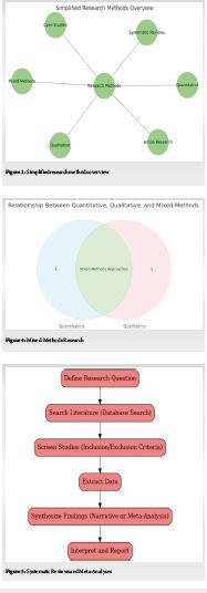
________________________________________
1. Quantitative Research Methods
Quantitative research emphasizes numerical data and statistical analysis. It is ideal for studies aiming to quantify relationships, test hypotheses, or derive generalizable patterns. Common quantitative approaches include:
• Surveys: Structured tools designed to collect data from large populations efficiently. These can be administered through various mediums such as online platforms, telephone interviews, or face-to-face interactions. Surveys excel in capturing trends, opinions, and behaviors but require careful design to avoid bias and ensure reliability.
• Experiments: Controlled investigations that manipulate variables to establish cause-and-effect relationships. These studies often utilize control and experimental groups, ensuring valid comparisons. Rigorous design, including randomization and blinding, enhances the reliability of results.
• Longitudinal Studies: Observational research conducted over extended periods to analyze changes and trends. For example, tracking health outcomes in a cohort over decades provides insights into disease progression and risk factors.
Quantitative methods are particularly powerful in disciplines such as economics, epidemiology, and education, where robust, objective data is paramount. However, their reliance on structured data collection instruments requires meticulous planning to ensure accuracy and relevance.
________________________________________
2. Qualitative Research Methods
Qualitative research focuses on understanding phenomena through non-numerical data such as words, images, or behaviors. It seeks to uncover deep insights into motivations, perceptions, and social dynamics. Prominent qualitative techniques include:
• Interviews: In-depth discussions with participants, ranging from structured formats to free-flowing conversations. These allow researchers to probe personal experiences, capturing rich, detailed narratives.
• Focus Groups: Interactive group discussions that explore collective opinions or behaviors. The dynamic nature of focus groups facilitates the emergence of diverse perspectives.
• Ethnography: Immersive studies that observe cultural or social practices within their natural context. Ethnographers often integrate themselves into communities, enabling a holistic understanding of social dynamics over time.
This method excels in fields like sociology, anthropology, and psychology, where exploring human experiences and cultural phenomena is vital. While qualitative research offers depth and nuance, it demands careful consideration of researcher bias and interpretation.
________________________________________
3. Mixed-Methods Research
Mixed-methods research combines the strengths of quantitative and qualitative approaches, enabling a more comprehensive understanding of research questions. Approaches include:
• Sequential Design: Conducting one type of research (e.g., quantitative) followed by the other (e.g., qualitative) to enrich findings.
• Concurrent Design: Simultaneously collecting quantitative and qualitative data to provide complementary insights.
• Embedded Design: Emphasizing one approach while using the other as a supplementary tool.
For example, a study on educational outcomes might use surveys to gather broad trends and follow up with interviews to explore underlying factors in detail. Mixed methods are invaluable in interdisciplinary research, addressing complex questions that require diverse perspectives.
 ________________________________________
4. Systematic Reviews and Meta-Analyses
These methods synthesize existing research, offering comprehensive insights:
• Systematic Reviews: Rigorous evaluations of all relevant studies on a specific topic. Researchers follow strict inclusion criteria to ensure unbiased and thorough analysis.
• Meta-Analyses: Statistical techniques that aggregate data from multiple studies to identify overarching trends. This method increases statistical power and clarifies conflicting findings across individual studies.
These approaches are pivotal in evidence-based disciplines like medicine, where decisions often rely on the aggregation of high-quality evidence. By identifying gaps and inconsistencies in existing research, they also guide future investigations.
________________________________________
5. Case Studies
Case studies offer an in-depth exploration of a single entity, event, or phenomenon. This method provides a detailed understanding of unique or complex cases, such as a groundbreaking technological innovation or a rare medical condition.
Researchers often combine various data collection techniques—interviews, observations, and archival analysis—to construct comprehensive narratives. While case studies yield rich insights, their findings are typically specific and may not be generalizable.
In fields like business, law, and clinical psychology, case studies serve as practical exemplars, providing valuable lessons and strategies for application.
________________________________________
6. Action Research
Action research integrates inquiry and practical application, emphasizing iterative cycles of reflection and improvement. It is particularly suited to professional environments, such as education and organizational management. The process involves:
1. Identifying a Problem: Collaborating with stakeholders to pinpoint an issue.
2. Planning an Intervention: Designing a strategy to address the problem.
3. Implementing the Plan: Acting on the strategy while monitoring its impact.
4. Reflecting and Revising: Analyzing results and refining approaches for future iterations.
Action research empowers participants, fostering collaboration and ensuring the research directly addresses real-world challenges. Its iterative nature facilitates continuous learning and improvement.
________________________________________
7. Observational Studies
Observational research involves studying subjects in their natural environments without manipulation. Key types include:
• Cross-Sectional Studies: Observations at a single point in time to identify patterns or correlations.
• Cohort Studies: Longitudinal studies tracking specific groups with shared characteristics. These are widely used in epidemiology to study disease progression or risk factors.
Observational studies are invaluable for generating hypotheses and understanding real-world behaviors. However, they require careful design to mitigate biases, as the lack of experimental control can complicate causal inferences.
________________________________________
Choosing the Right Method
Selecting an appropriate research method is a strategic decision influenced by:
• Research Goals: For hypothesis testing, quantitative methods may be optimal; for exploring complex behaviors, qualitative approaches may be preferable.
• Resources: Time, funding, and access to participants often dictate feasible methods.
• Ethical Considerations: Ensuring informed consent, confidentiality, and minimizing harm are paramount.
Scholars must also consider the balance between depth and breadth, often weighing the trade-offs of detailed exploration versus broad generalization.
________________________________________
Conclusion
The diversity of research methods reflects the complexity of the questions scholars aim to address. Each approach—from the precision of quantitative studies to the depth of qualitative insights—offers unique advantages and challenges. By carefully aligning their research questions, resources, and ethical considerations with their chosen method, researchers can generate findings that are not only robust but also transformative. Thoughtful selection and execution of research methods ultimately drive academic and societal progress, underscoring the enduring power of inquiry.
| How to Cite this Article: Kale S, Vatkar A, Shyam A, Hiwrale N, Gehlot O, Agarwal A. Exploring Different Methods of Study in Research: A Comprehensive Guide. Journal of Medical Thesis. 2024 July-December; 10(2):4-6. |

Full Text PDF | Full Text HTML
Crafting the Ideal Thesis: A Journey Toward Academic Excellence
Vol 10 | Issue 2 | July-December 2024 | page: 1-3 | Sachin Kale, Arvind Vatkar, Ashok Shyam, Nikhil Hiwrale, Ojasv Gehlot, Abhishek Agarwal
https://doi.org/10.13107/jmt.2024.v10.i02.234
Author: Sachin Kale [1], Arvind Vatkar [2], Ashok Shyam [3], Nikhil Hiwrale [1], Ojasv Gehlot [1], Abhishek Agarwal [1]
[1] Department of Orthopaedics, D Y Patil Hospital, Navi Mumbai, Maharashtra, India.
[2] Department of Orthopaedics, MGM Medical College, Navi Mumbai, Maharashtra, India.
[3] Department of Orthopaedics, Sancheti Institute for Orthopaedics and Rehabilitation, Pune, Maharashtra, India.
Address of Correspondence
Dr. Sachin Kale
Department of Orthopaedics, D Y Patil Hospital, Navi Mumbai, Maharashtra, India.
E-mail: sachinkale@gmail.com
Editorial
Introduction
The academic thesis represents the culmination of a scholar's intellectual journey, a work that encapsulates years of study, exploration, and analysis. It is not merely a document fulfilling a requirement but an artifact of scholarly rigor, creativity, and meticulous effort. Crafting an ideal thesis is a nuanced process, blending the art of storytelling with the precision of scientific inquiry. It involves an intricate balance of structure and innovation, where each component serves a distinct purpose in shaping a cohesive narrative. This editorial explores the critical elements of an ideal thesis in greater depth, offering a comprehensive guide to aspiring scholars.

The Pillars of an Ideal Thesis
1. Clear and Focused Research Question
The cornerstone of a strong thesis is a clearly defined research question or hypothesis. This element acts as the guiding star, shaping the study’s direction and ensuring coherence. A robust research question should be:
• Specific: Narrow enough to address particular aspects of a problem without being overly restrictive.
• Researchable: Feasible to investigate within the constraints of available time, resources, and data.
• Significant: Addressing a meaningful gap or unresolved issue in the field.
For instance, instead of broadly tackling "climate change impacts," a more focused question might be, "How does climate change affect agricultural yields in sub-Saharan Africa over the past two decades?" This clarity anchors the research, providing a clear trajectory and purpose. Scholars must invest substantial time in refining their research question, as this step determines the study’s scope and impact.
2. Comprehensive Literature Review
A literature review is more than a survey of existing studies; it is an intellectual synthesis that situates the thesis within the broader academic landscape. This section should achieve the following:
• Identify Key Frameworks: Highlight the seminal theories, models, and concepts that underpin the field.
• Critically Analyze Trends: Examine patterns, debates, and inconsistencies within existing research.
• Highlight Gaps: Identify unresolved questions or emerging areas of interest that the thesis seeks to address.
An effective literature review weaves these elements into a cohesive narrative, demonstrating the scholar’s command of the field and providing a foundation for their research. It also underscores the study’s originality, linking the research question to broader academic discourse. Instead of merely summarizing prior studies, the review should critique methodologies, identify contradictions, and justify the chosen focus.
3. Methodological Rigor
The methodology section serves as the research blueprint, detailing the "how" of the study. A well-executed methodology not only enhances the thesis’s credibility but also ensures its replicability. Key attributes of methodological rigor include:
• Appropriateness: Selecting methods that align with the research question, whether qualitative (e.g., interviews, ethnography), quantitative (e.g., surveys, experiments), or mixed methods.
• Justification: Providing a clear rationale for choosing specific methods, supported by theoretical and practical considerations.
• Transparency: Offering detailed descriptions of procedures, instruments, and analytical techniques to enable replication.
Acknowledging and addressing methodological limitations is equally critical. For instance, if data collection relies heavily on self-reported surveys, the researcher should discuss potential biases and how they were mitigated. This transparency reinforces the study’s validity and trustworthiness.
4. Original Contribution
Originality is the hallmark of a significant thesis, distinguishing it from existing work. Original contributions can take several forms:
• Empirical Data: Presenting novel findings derived from experiments, surveys, or fieldwork.
• Theoretical Innovation: Proposing new models, frameworks, or perspectives that challenge conventional paradigms.
• Practical Applications: Offering solutions to real-world problems, bridging the gap between theory and practice.
The thesis should articulate this originality explicitly, detailing how it advances the field or addresses a pressing societal issue. For instance, a thesis proposing a new method to enhance solar energy efficiency could highlight its implications for sustainable development and energy policy.
5. Logical Structure and Organization
An ideal thesis adheres to a logical structure that facilitates clarity and engagement. While formats may vary across disciplines, a common structure includes:
1. Introduction: Introducing the research question, objectives, significance, and context.
2. Literature Review: Establishing the theoretical and empirical background.
3. Methodology: Explaining the research design and processes.
4. Results: Presenting findings in a clear and systematic manner.
5. Discussion: Interpreting results, linking them to the research question and existing literature.
6. Conclusion: Summarizing insights, discussing implications, and suggesting future research directions.
Clear transitions between sections and a consistent narrative thread are essential, ensuring the thesis reads as a cohesive whole rather than a series of disconnected parts.
6. Engaging Writing Style
Effective communication is the bridge between academic rigor and accessibility. An ideal thesis achieves this balance by:
• Prioritizing Clarity: Avoiding unnecessary jargon and convoluted sentences.
• Embracing Conciseness: Expressing ideas succinctly while preserving depth.
• Utilizing Active Voice: Enhancing readability and immediacy.
• Maintaining Consistency: Adhering to a uniform tone and style throughout the document.
An engaging writing style not only enhances readability but also broadens the thesis’s impact, making it accessible to both specialists and non-specialists.
7. Thorough Analysis and Insightful Interpretation
The analysis transforms raw data into meaningful insights, forming the thesis’s intellectual core. Effective analysis involves:
• Alignment: Ensuring the analysis directly addresses the research objectives and question.
• Depth: Delving beyond surface-level observations to uncover deeper patterns and implications.
• Transparency: Clearly explaining the analytical techniques and processes used.
Interpretation is equally critical, involving:
• Connecting Findings: Relating results back to the research question and literature.
• Highlighting Implications: Discussing the broader significance of findings.
• Addressing Unexpected Outcomes: Exploring unanticipated results and their potential impact.
This dual focus on analysis and interpretation adds depth and richness to the thesis, showcasing the scholar’s intellectual agility.
8. Critical Reflection
An ideal thesis does not shy away from its limitations; instead, it embraces them as opportunities for future inquiry. Critical reflection includes:
• Discussing Constraints: Acknowledging the limitations of chosen methods and data.
• Identifying Biases: Addressing potential biases in data collection, analysis, or interpretation.
• Suggesting Future Research: Highlighting areas where further investigation is needed to build on the thesis’s findings.
This reflective approach demonstrates intellectual maturity, lending credibility and balance to the study.
9. Professional Presentation
The presentation of a thesis reflects the scholar’s attention to detail and professionalism. Essential elements include:
• Formatting: Adhering to institutional or publication guidelines for structure, citations, and layout.
• Proofreading: Ensuring the text is free of grammatical, typographical, and formatting errors.
• High-Quality Visuals: Incorporating clear and well-designed tables, graphs, and illustrations to support the narrative.
Professional presentation not only enhances readability but also underscores the scholar’s commitment to excellence.
10. Alignment with Ethical Standards
Ethical integrity is a cornerstone of credible scholarship. An ideal thesis upholds ethical standards by:
• Securing Permissions: Obtaining approvals for data collection and use of proprietary materials.
• Transparency: Clearly disclosing research processes, funding sources, and potential conflicts of interest.
• Avoiding Plagiarism: Ensuring all sources are properly attributed and cited.
Adherence to ethical standards safeguards the thesis’s credibility and contributes to the integrity of the academic community.
Conclusion
Crafting the ideal thesis is a demanding yet rewarding endeavor, one that requires meticulous planning, disciplined execution, and thoughtful reflection. By embracing the principles outlined above, scholars can produce theses that not only meet academic standards but also make meaningful contributions to their fields. Ultimately, an ideal thesis is more than a requirement; it is a legacy of intellectual excellence, a beacon for future research, and a testament to the scholar’s dedication to advancing knowledge.
| How to Cite this Article: Kale S, Vatkar A, Shyam A, Hiwrale N, Gehlot O, Agarwal A. Crafting the Ideal Thesis: A Journey Toward Academic Excellence. Journal of Medical Thesis. 2024 July-December ; 10(2): 1-3. |

Full Text PDF | Full Text HTML
Functional and Radiographic Predictors of Success in Transforaminal Lumbar Interbody Fusion: A Prospective Study of Lumbar Canal Stenosis
Vol 10 | Issue 2 | July-December 2024 | page: 48-52 | Abhishek Kothari, Chetan Pradhan, Atul Patil, Chetan Puram, Darshan Sonawane, Ashok Shyam, Parag Sancheti
https://doi.org/10.13107/jmt.2024.v10.i02.254
Author: Abhishek Kothari [1], Chetan Pradhan [1], Atul Patil [1], Chetan Puram [1], Darshan Sonawane [1], Ashok Shyam [1], Parag Sancheti [1]
[1] Department of Orthopaedics, Sancheti Institute of Orthopaedics and Rehabilitation, Pune, Maharashtra, India.
Address of Correspondence
Dr. Abhishek Kothari,
Sancheti institute for orthopedics and rehabilitation PG College, 16, Shivajinagar, pune -411005, Maharashtra
E-mail: dr.abhishekkothari@gmail.com
Abstract
Background: Transforaminal lumbar interbody fusion (TLIF) is commonly used to treat degenerative lumbar conditions that produce mechanical back pain, radiculopathy or instability. The procedure aims to decompress neural elements, restore disc height and provide segmental stability while limiting neural retraction. This study prospectively evaluates clinical and radiographic outcomes after TLIF in a tertiary centre, and examines how spinopelvic alignment change relates to patient-reported outcomes.
Methods: Consecutive patients with symptomatic lumbar canal stenosis and low-grade spondylolisthesis treated with TLIF between October 2019 and December 2021 were enrolled. Baseline assessment included VAS for back and leg pain, Oswestry Disability Index (ODI) and SF-36. Radiographs and MRI were used for preoperative planning; standing lateral films were used to calculate pelvic incidence (PI), pelvic tilt (PT), sacral slope (SS) and PI–LL mismatch. Standardised surgical techniques, postoperative care and follow-up at 6 and 12 months were used for outcome assessment.
Results: Of 48 enrolled, 40 patients completed one-year follow-up. Significant improvements were seen in VAS, ODI and SF-36 domains at one year. Many patients showed measurable correction of PI–LL mismatch. Clinical gains were frequent; however, the magnitude of radiographic change did not uniformly predict the degree of symptomatic improvement.
Conclusion: TLIF produced consistent clinical benefit at one year with low complication rates. While sagittal alignment correction often accompanied improved function, imaging gains alone did not guarantee greater symptomatic relief, highlighting the multifactorial nature of recovery after fusion.
Keywords: TLIF, Lumbar canal stenosis, Spondylolisthesis, Oswestry Disability Index, Spinopelvic alignment
Introduction:
Transforaminal lumbar interbody fusion (TLIF) has become a widely used technique for addressing degenerative lumbar disorders that produce neural compression, segmental instability or chronic mechanical back pain. TLIF permits direct posterior decompression while allowing placement of an interbody graft or cage through a unilateral transforaminal corridor, thereby reducing the need for extensive neural retraction that characterized earlier posterior interbody techniques. This technical advantage helped TLIF gain popularity as an option that balances safe neural decompression with restoration of anterior column support and segmental stability. [1-6]
Historically, surgeons relied on posterolateral fusion and posterior lumbar interbody fusion (PLIF) for many degenerative indications; however, PLIF involves greater bilateral neural manipulation and has been associated with certain approach-related risks. Modern outcome assessment emphasises patient-reported measures such as VAS, ODI and SF-36 because radiographic fusion alone does not fully capture the patient’s functional recovery and quality of life. At the same time, restoration of sagittal balance — often summarized by spinopelvic parameters such as pelvic incidence (PI), pelvic tilt (PT), sacral slope (SS) and PI–LL mismatch — has gained attention for its association with long-term function and adjacent-segment health. Surgeons now balance the goals of neural decompression and fusion with alignment objectives to optimise both short- and long-term outcomes. [2][4][6]
The available literature suggests TLIF is effective and broadly safe in selected patients, yet questions remain about how well radiographic correction predicts patient-centred improvement. This study was designed to prospectively evaluate clinico-radiological outcomes after TLIF in patients with lumbar canal stenosis and low-grade spondylolisthesis, and to explore the relationship between changes in spinopelvic parameters and clinical recovery. [1-6]
Review of Literature
Multiple comparative studies and meta-analyses have examined TLIF relative to other fusion strategies (PLIF, ALIF and posterolateral fusion). Overall, these syntheses report broadly similar fusion rates across interbody techniques but note differences in approach-related morbidity, operative times and the capacity to restore segmental lordosis. Several series show TLIF may carry a lower risk of neural retraction-related complications compared with PLIF while achieving comparable clinical outcomes and fusion efficacy. [7-13]
Randomized trials and cohort studies focusing on degenerative spondylolisthesis have shown that adding fusion to decompression can offer better medium-term improvements in pain and function for selected patients, although the balance between benefit and the risk of reoperation depends on careful patient selection. Not all patients with lumbar stenosis require fusion — the decision rests on instability, deformity, and individual symptom profiles. Meta-analyses highlight that minimally invasive variants of TLIF reduce blood loss and early morbidity while maintaining similar fusion and symptomatic results when compared with open techniques in appropriate hands. [7][8][9]
The role of sagittal balance and spinopelvic parameters in predicting outcomes after lumbar fusion has been increasingly studied. Excessive PI–LL mismatch and sagittal malalignment have been associated with worse health-related outcomes and a higher incidence of adjacent-segment disease in some series, prompting surgeons to incorporate alignment goals into surgical planning. However, multiple reports caution that radiographic correction alone does not uniformly translate into proportional improvements in pain or disability; clinical recovery is mediated by neurological status, chronicity of symptoms, paraspinal muscle condition, comorbidities and psychosocial factors. Thus, integrated assessment of clinical and radiographic outcomes is essential when evaluating the true benefit of fusion procedures. [14- 20]
Materials and Methods
This single-centre prospective study enrolled consecutive patients who underwent TLIF for symptomatic lumbar canal stenosis with or without low-grade degenerative spondylolisthesis from October 2019 through December 2021. Institutional ethics approval and written informed consent were obtained. Inclusion criteria were age >20 years, clinical and MRI confirmation of neural compression and failure of conservative care (physiotherapy and medication) for at least six weeks. Exclusion criteria included active spinal infection, metastatic disease, high-grade spondylolisthesis, severe osteoporosis and inability to comply with follow-up.
Preoperative evaluation included detailed clinical assessment, neurological examination, VAS for back and leg pain, ODI and SF-36. Imaging included standing AP and lateral radiographs incorporating femoral heads (for spinopelvic measurements), flexion–extension films and MRI for neural element assessment. Spinopelvic parameters measured on standing lateral films included pelvic incidence (PI), pelvic tilt (PT), sacral slope (SS) and lumbar lordosis (LL) to calculate PI–LL mismatch. Baseline comorbidities, smoking status and medication history were recorded.
Surgical technique: All procedures were performed under general anaesthesia via a midline posterior approach. Unilateral facetectomy and foraminotomy were performed on the symptomatic side to decompress nerve roots. Discectomy and endplate preparation were followed by insertion of an interbody cage packed with local autograft; pedicle screw fixation was applied bilaterally and rods contoured to achieve segmental correction. Fluoroscopic guidance confirmed implant position. Perioperative antibiotics and standard thromboprophylaxis were used.
Postoperative care included early mobilisation, drain removal as per output, staged physiotherapy and outpatient follow-up at 6 and 12 months. Outcome analysis compared preoperative and follow-up VAS, ODI and SF-36 scores using paired tests; radiographic changes in spinopelvic parameters were evaluated, and correlations between radiographic and clinical changes explored. Statistical significance was set at p<0.05.
Results
Forty-eight patients were enrolled; 40 completed one-year follow-up and formed the analysis cohort. The mean age was 52 years (range 38–72) with a modest female predominance. The majority underwent single-level fusion, most commonly at L4–L5. Common comorbidities included well-controlled hypertension and type-2 diabetes in a subset. Preoperative neurological deficits were present in several patients; most showed partial or full recovery by one year.
Clinical outcomes: Median VAS back pain decreased from 7 (preop) to 1 at one year; leg pain VAS showed a similar fall. Mean ODI improved markedly from roughly 68% preoperatively to about 18% at one year, indicating substantial reduction in disability. SF-36 physical and bodily pain domains improved significantly across the cohort.
Radiographic outcomes: Standing lateral films at one year demonstrated measurable changes in spinopelvic parameters in many patients, with a general tendency toward reduction in PI–LL mismatch after fusion. Complications were infrequent and consistent with published TLIF series: a small number of transient dural tears, one case of superficial wound infection managed conservatively, and no perioperative mortalities. Overall, the majority of patients achieved meaningful clinical improvement at one year.
Discussion
In this series, patients treated with TLIF for lumbar canal stenosis and low-grade spondylolisthesis experienced clear and sustained improvement in pain, disability and quality of life at one year. The reductions in VAS scores and the marked fall in ODI mirror findings reported in other clinical series, supporting TLIF’s role in achieving neural decompression, restoring disc height and providing segmental stability that together reduce mechanical back pain and radicular symptoms. [14][15]
Radiographs showed that many patients had measurable improvement in sagittal parameters, particularly a reduction in PI–LL mismatch. Restoration of a more favourable sagittal alignment is encouraging because several studies associate better alignment with improved long-term function and a lower risk of adjacent-segment problems. However, we also observed that imaging and symptoms do not always move in lockstep: several patients reported large functional gains despite only modest radiographic change, and a few with good radiographic correction reported only modest symptomatic relief. This mismatch highlights that radiographic correction is important but not by itself determinative of patient experience. [18][19][20]
There are several plausible reasons for this discordance. Duration and chronicity of preoperative symptoms, severity of preoperative neurological deficit, condition of the paraspinal muscles, and patient comorbidities (for example diabetes or peripheral neuropathy) influence neural recovery and pain perception. Psychosocial factors and expectations also shape reported outcomes, as do rehabilitation and return-to-activity practices after surgery. Classic descriptions of the multifactorial nature of low back pain remind us that structural correction addresses a single domain within a broader biopsychosocial context. [16][17]
Clinically, these observations suggest a balanced approach: aim for reasonable sagittal correction during fusion, but prioritise individualized goals based on the patient’s overall health, symptom history and functional needs rather than pursuing perfect radiographic numbers alone. Careful patient selection, meticulous surgical technique, prompt mobilisation and a structured rehabilitation programme appear to contribute substantially to favourable recovery. Limitations of our study include single-centre design, modest sample size and one-year follow-up, which constrain assessment of long-term fusion durability and late adjacent-level disease. Larger, longer studies would help identify which preoperative and intraoperative factors most reliably predict when radiographic improvement will translate into durable, patient-centred benefit. [18][19][20]
Conclusion
TLIF produced consistent and meaningful improvements in pain, disability and quality of life at one year in this cohort of patients with lumbar canal stenosis and low-grade spondylolisthesis. Radiographic correction of sagittal alignment often accompanied clinical gains, yet imaging improvement alone did not guarantee a larger symptomatic benefit for every patient. Recovery after TLIF is multifactorial: careful patient selection, realistic and individualized alignment goals, meticulous surgical technique, and a structured rehabilitation plan are all important contributors to favourable outcomes. The low complication rates observed suggest TLIF is a reliable option in experienced hands. Further research with larger cohorts and longer follow-up is needed to better characterise predictors of sustained clinical benefit and the long-term relationship between alignment, fusion status and adjacent-segment health.
References
1. Resnick DK, Choudhri TF, Dailey AT, Groff MW, Khoo L, Matz PG, et al. Guidelines for the performance of fusion procedures for degenerative disease of the lumbar spine. Part 7: intractable low-back pain without stenosis or spondylolisthesis. J Neurosurg Spine. 2005 Jun; 2(6):670–2.
2. Eck JC, Hodges S, Humphreys SC. Minimally invasive lumbar spinal fusion. J Am Acad Orthop Surg. 2007 Jun; 15(6):321–9.
3. Mobbs RJ, Sivabalan P, Li J. Minimally invasive surgery compared to open spinal fusion for the treatment of degenerative lumbar spine pathologies. J Clin Neurosci. 2012 Jun; 19(6):829–35.
4. Zhao J, Zhang S, Li X, He B, Ou Y, Jiang D. Comparison of Minimally Invasive and Open Transforaminal Lumbar Interbody Fusion for Lumbar Disc Herniation: A Retrospective Cohort Study. Med Sci Monit. 2018 Dec 1; 24:8693–8.
5. Mobbs RJ, Phan K, Malham G, Seex K, Rao PJ. Lumbar interbody fusion: techniques, indications and comparison of interbody fusion options including PLIF, TLIF, MI-TLIF, OLIF/ATP, LLIF and ALIF. J Spine Surg. 2015 Dec;1(1):2–18.
6. Uçar BY, Özcan Ç, Polat Ö, Aman T. Transforaminal Lumbar Interbody Fusion For Lumbar Degenerative Disease: Patient Selection And Perspectives. Orthop Res Rev. 2019 Nov; 11:183–9.
7. Macki M, Bydon M, Weingart R, Sciubba D, Wolinsky J-P, Gokaslan ZL, et al. Posterolateral fusion with interbody for lumbar spondylolisthesis is associated with less repeat surgery than posterolateral fusion alone. Clin Neurol Neurosurg. 2015 Nov; 138:117–23.
8. Lan T, Hu S-Y, Zhang Y-T, Zheng Y-C, Zhang R, Shen Z, et al. Comparison Between Posterior Lumbar Interbody Fusion and Transforaminal Lumbar Interbody Fusion for the Treatment of Lumbar Degenerative Diseases: A Systematic Review and Meta-Analysis. World Neurosurg. 2018 Apr; 112:86–93.
9. Harms JG, Jeszenszky D. Die posteriore, lumbale, interkorporelle Fusion in unilateraler transforaminaler Technik. Oper Orthop Traumatol. 1998 Jun 1; 10(2):90–102.
10. de Kunder SL, van Kuijk SMJ, Rijkers K, Caelers IJMH, van Hemert WLW, de Bie… [Entry continues in dissertation].
11. Yavin D, et al. [Systematic review/meta-analysis on comparative safety and efficacy of lumbar fusion, decompression alone or nonoperative care]. Spine J. 2017.
12. Ghogawala Z, et al. Laminectomy plus fusion versus laminectomy alone for symptomatic lumbar grade I degenerative spondylolisthesis: randomized trial. 2016.
13. Frymoyer JW, Pope MH, Clements JH, Wilder DG, MacPherson B, Ashikaga T. Risk factors in low-back pain. J Bone Joint Surg Am. 1983 Feb; 65(2):213–8.
14. Dillane JB, Fry J, Kalton G. Acute back syndrome—a study from general practice. Br Med J. 1966 Jul 9; 2(5505):82–4.
15. Kelsey JL, White AA. Epidemiology and impact of low-back pain. Spine. 1980 Apr; 5(2):133–42.
16. Kirkaldy-Willis WH, Wedge JH, Yong-Hing K, Reilly J. Pathology and pathogenesis of lumbar spondylosis and stenosis. Spine. 1978 Dec; 3(4):319–28.
17. Mixter WF, Barr JS. Rupture of the Intervertebral Disc and Its Association with Lumbar and Leg Pain. N Engl J Med. 1934.
18. Mac-Thiong J-M, Wang Z, de Guise JA, Labelle H. Postural Model of Sagittal Spino-Pelvic Alignment and Its Relevance for Lumbosacral Developmental Spondylolisthesis. Spine. 2008 Oct; 33(21):2316–25.
19. Le Huec JC, Charosky S, Barrey C, Rigal J, Aunoble S. Sagittal imbalance cascade for simple degenerative spine and consequences: algorithm of decision for appropriate treatment. Eur Spine J. 2011 Sep; 20(S5):699–703.
20. Barrey C, Roussouly P, Perrin G, Le Huec J-C. Sagittal balance disorders in severe degenerative spine. Eur Spine J. 2011 Sep; 20(S5):626–33.
| How to Cite this Article: Kothari A, Pradhan C, Patil A, Puram C, Sonawane D, Shyam A, Sancheti P. Functional and Radiographic Predictors of Success in Transforaminal Lumbar Interbody Fusion: A Prospective Study of Lumbar Canal Stenosis. Journal of Medical Thesis. 2024 July-December; 10(2): 48-51. |
Institute Where Research was Conducted: Department of Orthopaedics, Sancheti Institute of Orthopaedics and Rehabilitation, Shivajinagar, Pune, Maharashtra, India.
University Affiliation: Maharashtra University of Health Sciences (MUHS), Nashik, Maharashtra, India
Year of Acceptance of Thesis: 2019
Full Text HTML | Full Text PDF

Evaluating the Impact of Anatomical Restoration of Hip Offset and Leg Length on One Year THR Outcomes
Vol 10 | Issue 2 | July-December 2024 | page: 44-47 | Pavan Patil, Rajeev Joshi, Sahil Sanghavi, Mahavir Dugad, Darshan Sonawane, Ashok Shyam, Parag Sancheti
https://doi.org/10.13107/jmt.2024.v10.i02.252
Author: Pavan Patil [1], Rajeev Joshi [1], Sahil Sanghavi [1], Mahavir Dugad [1], Darshan Sonawane [1], Ashok Shyam [1], Parag Sancheti [1]
[1] Department of Orthopaedics, Sancheti Institute of Orthopaedics and Rehabilitation, Pune, Maharashtra, India.
Address of Correspondence
Dr. Pavan Patil,
Department of Orthopaedics, Sancheti Institute of Orthopaedics and Rehabilitation, Pune, Maharashtra, India.
E-mail: drpavan010@gmail.com
Abstract
Background: Restoring femoral offset and limb length is essential for pain relief, stable gait and patient satisfaction after primary total hip replacement. Small deviations can alter abductor mechanics and affect function.
Methods: We prospectively studied 150 patients undergoing primary total hip replacement at a single centre from October 2019 to December 2021. Preoperative templating used standardized AP pelvic radiographs. Leg-length discrepancy was measured by the Woolson inter-teardrop method and global offset was calculated from acetabular and femoral measurements. Clinical outcomes were recorded before surgery and at postoperative intervals. Data were analysed using appropriate statistical tests for repeated measures.
Results: Most patients achieved postoperative limb-length and global-offset differences within five millimeters of the contralateral hip. Pain scores fell and functional scores rose consistently over the first postoperative year. Radiographic review showed stable components and a low complication rate.
Conclusion: Careful radiographic templating combined with attentive intraoperative technique can reliably restore offset and limb length within clinically acceptable limits in routine primary total hip replacement. This reconstruction is therefore associated with meaningful early gains in pain relief, mobility and quality of life; longer follow-up will clarify effects on implant survivorship.
Keywords: Total hip replacement, Femoral offset, Leg-length discrepancy, Templating, Patient-reported outcomes, Radiographic measurement
Introduction:
Total hip replacement is one of orthopedics’ most successful procedures and has transformed the lives of many patients with end-stage hip disease. To achieve predictable relief of pain and recovery of function, surgeons must do more than insert well-fixed implants — they must also restore the hip’s native geometry so that muscles and soft tissues work efficiently. Restoring femoral offset and the correct limb length are central to rebuilding a balanced hip: femoral offset determines the abductor lever arm and thus affects muscle strength and joint reaction forces, while leg-length inequality can cause limp, back pain and dissatisfaction if not corrected. [1–5]
Small changes in offset or length can influence abductor tension, gait mechanics and patient perception. Decreasing offset tends to shorten the abductor lever arm and can reduce muscular efficiency; increasing offset beyond what is necessary may over-tension soft tissues and cause discomfort. Likewise, even modest leg-length differences can be noticeable to patients, and larger discrepancies frequently lead to symptomatic complaints. Preoperative templating on standardized radiographs and careful intraoperative assessment are practical tools to reduce these risks in routine practice. This study used those methods to examine whether restoration of offset and limb length correlates with better patient-reported outcomes and to evaluate the reliability of plain radiographic measurement in a clinical setting. [1–5]
Review of literature
Earlier biomechanical and clinical work has repeatedly highlighted the importance of femoral offset and limb length in hip arthroplasty. Several studies showed that changes in offset alter abductor mechanics and can translate into measurable differences in gait and muscle strength. Restoration of offset has been associated with improved abductor leverage and, in some series, lower rates of component-related problems. [6–9]
Radiographic templating on AP pelvis films is the standard approach in most centres because it is accessible and practical, though it has recognized limitations: plain radiographs can underestimate true three-dimensional offset when compared with CT and are sensitive to pelvic rotation and magnification. Where available, 3-D planning provides greater accuracy, but for routine primary cases standardized radiography combined with careful technique offers a pragmatic balance of accuracy and convenience. [6–10]
Clinical thresholds vary between studies: some reports suggest that length differences up to about 10–20 mm may be tolerated by many patients, while others note that discrepancies above roughly 5–10 mm are more likely to affect gait symmetry or satisfaction. Gait-analysis studies indicate that combined deviations of length and offset correlate with altered kinematics; however, the effect on patient-reported outcome measures is often modest when deviations are small. Taken together, the literature supports aiming to restore offset and limb length as closely as practical while recognizing that small deviations within accepted limits frequently have limited clinical impact. [11–13]
Materials and Methods
This single-centre prospective study included 150 consecutive patients who underwent primary total hip replacement from October 2019 to December 2021. Exclusion criteria were congenital hip deformity, bilateral symptomatic disease at presentation, ankylosed hips and arthroplasty for acute trauma. Baseline demographic data and preoperative scores (Harris Hip Score, WOMAC, Oxford Hip Score, VAS and SF-36) were recorded.
Preoperative templating was performed on standardized AP pelvic radiographs. Leg length was measured by the inter-teardrop to lesser trochanter distance (Woolson method) and global offset as the sum of acetabular and femoral offsets on the AP film. To reduce radiographic error we used consistent patient positioning and magnification markers. Surgeries were performed predominantly via a posterolateral approach with templated osteotomy and trial reductions to confirm stability and soft-tissue balance. Modular head/neck options were used when intraoperative adjustments were needed.
Postoperative radiographs and clinical evaluations were performed at 6 weeks, 6 months and 12 months. Radiographic assessment included component position and signs of loosening or osteolysis. Continuous variables are presented as medians (IQR) and categorical data as counts (percentages). Repeated measures were analysed with linear mixed-effects models to account for within-patient correlations over time.
Results
The results reflect a large, generally healthy cohort of 151 patients (median age 51 years, IQR 39–60; 68% male). Median hospital stay was 5 days; median height 160 cm, weight 67 kg and BMI 25.7. Comorbidities were present in 58% (hypertension 32%, diabetes 13%, hypothyroid 7%, ischemic heart disease 5%). Radiographically, median global offset on the operated side was 7.24 cm (IQR 6.95–7.68) versus 7.24 cm (IQR 6.83–7.93) on the contralateral side (p = 0.52), while median leg-length measured 4.51 cm (IQR 4.06–4.99) operated versus 4.55 cm (IQR 3.90–4.89) contralateral (p < 0.001), indicating only small, clinically acceptable differences for most patients. Patient-reported outcomes improved markedly from baseline to 12 months: Harris Hip Score median 34 → 84, WOMAC 62 → 12, Oxford Hip Score 15 → 42, VAS pain 8 → 1, and SF-36 physical function 27 → 80 (all trends p < 0.0001). At one year median pain was low (VAS 1, IQR 1–2) and quality-of-life domains rose substantially. Early complications were uncommon: there were no cups with progressive migration at final review and only a few osteolytic lesions. Patients with offset or length differences within small thresholds (approximately ≤5 mm) experienced functional gains comparable to the whole cohort; larger discrepancies were rare and therefore made subgroup analysis limited.
Discussion
This series confirms that careful preoperative templating and attentive intraoperative technique can restore hip geometry — offset and limb length — within clinically acceptable margins for most patients undergoing primary THR. The findings echo prior biomechanical and clinical studies that link accurate offset reconstruction to improved abductor mechanics and suggest that restoring anatomy supports better objective and subjective outcomes. [14–16]
The literature describes a range of acceptable thresholds for offset and length discrepancies. While some degree of variation may be tolerated, deviations above commonly cited thresholds are more likely to alter gait kinematics and provoke symptoms. Where offset cannot be restored by a standard implant configuration, modular components or high-offset stems provide practical intraoperative options to fine-tune reconstruction. Radiographic templating on AP films remains a pragmatic method for routine cases; although CT-based planning is more precise, it is not necessary for every primary THR and is best reserved for complex anatomy or dysplastic hips. [16–18]
Several larger cohort and systematic reviews report similar conclusions: small deviations from native offset or limb length often have little effect on PROMs, but under-correction of offset and larger leg-length inequality are associated with worse functional measures and gait abnormalities in some studies. Surgeons should therefore aim for accurate restoration while recognizing that small residual differences are common and frequently clinically acceptable. [19–20]
Conclusion
In this cohort of 150 primary THR patients, restoration of limb length and global offset to within small, clinically acceptable margins was achieved in the majority. These patients showed consistent improvement in pain, function and health-related quality of life at one year. Standardized radiographic templating and intraoperative verification provide an effective, pragmatic approach to biomechanical reconstruction in routine primary THR. Three-dimensional planning remains valuable in selected complex cases.
References
1. Learmonth ID, Young C, Rorabeck C. The operation of the century: total hip replacement. Lancet. 2007; 370:1508–1519.
2. Bjordal F, Bjorgul K. The role of femoral offset and abductor lever arm in total hip arthroplasty. J Orthop Traumatol. 2015; 16(4):325–330.
3. Parry MC, Povey J, Blom AW, Whitehouse MR. Comparison of acetabular bone resection, offset, leg length and postoperative function between hip resurfacing arthroplasty and total hip arthroplasty. J Arthroplast. 2015; 30(10):1799–1803.
4. Hassani H, Cherix S, Ek ET, Rudiger HA. Comparisons of preoperative three-dimensional planning and surgical reconstruction in primary cementless total hip arthroplasty. J Arthroplast. 2014; 29(6):1273–1277.
5. Asayama I, Chamnongkich S, Simpson KJ, Kinsey TL, Mahoney OM. Reconstructed hip joint position and abductor muscle strength after total hip arthroplasty. J Arthroplast. 2005; 20(4):414–420.
6. Cassidy KA, Noticewala MS, Macaulay W, Lee JH, Geller JA. Effect of femoral offset on pain and function after total hip arthroplasty. J Arthroplast. 2012; 27(10):1863–1869.
7. Mahmood SS, Mukka SS, Crnalic S, Wretenberg P, Sayed-Noor AS. Association between changes in global femoral offset after total hip arthroplasty and function, quality of life, and abductor muscle strength: a prospective cohort study. Acta Orthop. 2016; 87(1):36–41.
8. Spalding TJ. Effect of femoral offset on motion and abductor muscle strength after total hip arthroplasty. J Bone Joint Surg Br. 1996; 78:997–998.
9. Lecerf G, Fessy MH, Philippot R, Massin P, Giraud F, Flecher X, et al. Femoral offset: anatomical concept, definition, assessment, implications for preoperative templating and hip arthroplasty. Orthop Traumatol Surg Res. 2009; 95(3):210–219.
10. Benedetti MG, Catani F, Benedetti E, Berti L, Di Gioia A, Giannini S. To what extent does leg length discrepancy impair motor activity in patients after total hip arthroplasty? Int Orthop. 2010; 34(8):1115–1121.
11. Plaass C, Clauss M, Ochsner PE, Ilchmann T. Influence of leg length discrepancy on clinical results after total hip arthroplasty—A prospective clinical trial. Hip Int. 2011; 21(4):441–449.
12. Desai AS, Dramis A, Board TN. Leg length discrepancy after total hip arthroplasty: a review of literature. Curr Rev Musculoskelet Med. 2013; 6(4):336–341.
13. Sariali E, Klouche S, Mouttet A, Pascal-Moussellard H. The effect of femoral offset modification on gait after total hip arthroplasty. Acta Orthop. 2014; 85(2):123–127.
14. Zhang Y, He W, and Cheng T, Zhang X. Total hip arthroplasty: leg length discrepancy affects functional outcomes and patient’s gait. Cell Biochem Biophys. 2015; 72(1):215–219.
15. Li J, McWilliams AB, Jin Z, Fisher J, Stone MH, Redmond AC, Stewart TD. Unilateral total hip replacement patients with symptomatic leg length inequality have abnormal hip biomechanics during walking. Clin Biomech. 2015; 30(5):513–519.
16. Renkawitz T, Weber T, Dullien S, Woerner M, Dendorfer S, Grifka J, Weber M. Leg length and offset differences above 5 mm after total hip arthroplasty are associated with altered gait kinematics. Gait Posture. 2016; 49:196–201.
17. Flecher X, Ollivier M, Argenson JN. Lower limb length and offset in total hip arthroplasty. Orthop Traumatol Surg Res. 2016; 102(1 Suppl):S9–S20.
18. Bolink SA, Lenguerrand E, Brunton LR, Hinds N, Wylde V, Heyligers IC, et al. The association of leg length and offset reconstruction after total hip arthroplasty with clinical outcomes. Clin Biomech. 2019; 68:89–95.
19. Shapira J, Chen SL, Rosinsky PJ, Maldonado DR, Meghpara M, Lall AC, Domb BG. The effect of postoperative femoral offset on outcomes after hip arthroplasty: A systematic review. J Orthop. 2020; 22:5–11.
20. Hassani H, Cherix S, Ek ET, Rudiger HA. Comparisons of preoperative three-dimensional planning and surgical reconstruction in primary cementless total hip arthroplasty. J Arthroplast. 2014; 29(6):1273–1277.
| How to Cite this Article: Patil P, Joshi R, Sanghavi S, Dugad M, Sonawane D, Shyam A, Sancheti P. Evaluating the Impact of Anatomical Restoration of Hip Offset and Leg Length on One-Year THR Outcomes ournal of Medical Journal Medical Thesis. 2024 July-December; 10(2): 44-47. |
Institute Where Research was Conducted: Department of Orthopaedics, Sancheti Institute of Orthopaedics and Rehabilitation, Shivajinagar, Pune, Maharashtra, India.
India.
University Affiliation: Maharashtra University Of Health Sciences (MUHS), Nashik,
Maharashtra, India
Year of Acceptance of Thesis: 2019
Full Text HTML | Full Text PDF 
Clinical and Functional Outcomes of Limb Reconstruction System in Infected Non-Union of the Femur: A Prospective-Retrospective Cohort Study
Vol 10 | Issue 2 | July-December 2024 | page: 40-43 | Jenil Patel, Rajeev Joshi, Sahil Sanghavi, Mahavir Dugad, Darshan Sonawane, Ashok Shyam, Parag Sancheti
https://doi.org/10.13107/jmt.2024.v10.i02.250
Author: Jenil Patel, Rajeev Joshi, Sahil Sanghavi, Mahavir Dugad, Darshan Sonawane, Ashok Shyam, Parag Sancheti
[1] Department of Orthopaedics, Sancheti Institute of Orthopaedics and
Rehabilitation, Pune, Maharashtra, India.
Address of Correspondence
Dr. Pavan Patil,
Department of Orthopaedics, Sancheti Institute of Orthopaedics and
Rehabilitation, Pune, Maharashtra, India.
E-mail: drpavan010@gmail.com
Abstract
Background: Infected non-union of the femur causes prolonged pain, disability and loss of livelihood. Treating infection while restoring bone continuity, length and alignment is challenging. The Limb Reconstruction System (LRS) is a uniplanar external fixator that can address bone loss, deformity and infection together.
Methods: This combined prospective-retrospective series included adult patients treated June 2018–June 2022. After radical debridement and culture-directed antibiotics, LRS was used according to defect size: monofocal compression for small defects, bone transport for larger gaps and bifocal techniques when required. Patients followed a protocol of pin-site care, regular radiographs, clinical review and physiotherapy. Recorded outcomes were time to union, infection control, ASAMI scores and limb length discrepancy.
Results: Twenty-one patients were treated. Twenty achieved radiological union; infection was controlled in 18. Most patients regained useful function with good or excellent ASAMI scores. Residual shortening was under 2 cm and managed conservatively..
Conclusion: When combined with thorough debridement and structured rehabilitation, LRS yields high union rates and acceptable function in many infected femoral non-unions. Vigilant pin care and engagement are essential.
Keywords: Infected non-union, Femur, Limb Reconstruction System, Bone transport, External fixation
Introduction
Infected non-union of the femur is a serious and often devastating complication after fracture. The combination of infection and failure of bone healing prolongs disability, interferes with daily life and places heavy social and economic burdens on patients and families. Management is challenging because one must eradicate infection, restore bone continuity and alignment, correct limb length discrepancy, and preserve joint function — sometimes all at once. Early recognition of the factors that favour non-union, such as high-energy injury, soft-tissue damage, comminution and prior failed fixation, helps plan the reconstruction approach. Radical debridement to remove devitalized bone and infected tissue is the cornerstone of treatment; without it, eradication of infection is unlikely and any reconstruction risks failure. The tension-stress principles described for distraction osteogenesis revolutionized reconstructive options and made staged bone transport and lengthening feasible alternatives to bone grafting for large defects. In practice, circular ring fixators based on those principles are effective but can be unwieldy for the femur because of their bulk and the difficulty of fitting them to the thigh. To reduce these disadvantages, monolateral systems such as the Limb Reconstruction System (LRS) and related dynamic axial or monorail devices were developed. These systems allow bone transport, compression-distraction and acute or gradual deformity correction using a simpler, lighter frame that is often better tolerated by patients. Contemporary series report acceptable union and infection control rates with monolateral constructs when radical debridement, appropriate antibiotic therapy and careful follow-up are applied. In this work we report our experience with the LRS for infected femoral non-union, focusing on union rates, infection eradication, limb length restoration, alignment and functional recovery. [1-7]
Review of literature
External fixation has evolved substantially since its early introductions, and its role in contaminated wounds and infected non-union is well established. Early unilateral and dynamic axial fixators showed the benefits of preserving soft-tissue access while enabling stability and early weight bearing. The Ilizarov method, grounded in the tension-stress effect and distraction osteogenesis, remains a central technique for reconstructing bone loss, correcting deformity and managing infection simultaneously. While effective, ring fixators have recognized disadvantages including a steep learning curve, bulkiness, patient discomfort and periodical pin-care challenges. These limitations prompted the development and refinement of monolateral devices and limb reconstruction rails that permit uniplanar transport and lengthening with a lower profile, simpler application and improved patient comfort. Comparative reports indicate that, in selected femoral reconstructions, monolateral systems can achieve outcomes comparable to circular frames in terms of union and infection control while reducing hardware complexity. The key surgical strategy across studies emphasizes aggressive excision of necrotic bone and soft tissue, obtaining deep cultures, using targeted systemic antibiotics and planning reconstruction based on defect size: one-stage grafting for small defects, antibiotic nails for some intramedullary infections and bone transport for larger segmental losses. Reviews also stress the importance of multidisciplinary care, diligent pin-site protocols and sustained physiotherapy to manage stiffness and maintain limb function. Evidence from recent monolateral series supports the LRS as a versatile tool for femoral reconstruction — permitting monofocal or bifocal lengthening, acute correction where needed, and staged bone transport when defects are significant. These reports highlight acceptable union rates and functional gains when careful patient selection and meticulous technique are employed. [8- 20]
Methods and Materials
This combined prospective and retrospective study included patients treated for infected femoral non-union with the Limb Reconstruction System (LRS) at our tertiary centre between June 2018 and June 2022. Adults aged 18–70 years with clinical, radiological and microbiological evidence of infected non-union were included after informed consent; excluded were skeletally immature patients, tuberculous non-union and cases with severe neurological impairment of the affected limb. Preoperative evaluation comprised detailed history (initial injury, prior surgeries, duration of non-union), physical examination (sinuses, drainage, deformity, shortening and joint range), laboratory tests (Hb, leukocyte count, ESR, CRP) and deep tissue cultures obtained at debridement. Radiographs (AP and lateral) defined defect size, alignment and bone quality. Operative strategy began with aggressive debridement and excision of all devitalized bone and soft tissue until healthy bleeding margins were achieved. The LRS was then applied in a configuration suited to the defect: monofocal compression for non-unions with minimal bone loss, bone transport for moderate to large segmental defects, and bifocal constructs where simultaneous docking and distraction were needed. Acute shortening followed by later lengthening was used in select cases to diminish soft-tissue tension. Postoperatively patients were instructed in pin-site care and commenced early mobilisation; distraction followed standard callotasis protocols with radiographs every two weeks during distraction and monthly during consolidation. Outcomes recorded were time to clinical and radiological union, infection eradication, ASAMI bone and functional scores, final limb length discrepancy, alignment and complications including pin-tract infection, pin loosening and joint stiffness. Follow-up extended for a minimum period suited to consolidation in each case. [15- 17]
Results
Twenty-one patients met the inclusion criteria. Patient ages ranged from 19 to 51 years (mean ~36 years) with strong male predominance. Right femur was involved in most cases. A majority of the cohort had sustained open fractures initially and had undergone prior operative fixation. After radical debridement and application of the LRS, 20 of 21 patients achieved clinical and radiological union; a single case required further intervention for persistent non-union. Infection was eradicated in 18 patients while three had persistent or recurrent infection associated with resistant organisms and required additional surgical or medical management. ASAMI bone results were mostly excellent or good, and functional scores reflected satisfactory recovery in the majority. Average residual limb length discrepancy at final follow-up was under 2 cm in all patients and was managed conservatively when necessary. Notable complications were knee stiffness in several patients, hip stiffness in some, limb strength asymmetry and pin-site problems including superficial infections and occasional pin loosening; most complications were managed non-operatively or with minor procedures. Overall the LRS provided stable, versatile fixation that allowed bone transport or compression while supporting early mobilisation and functional rehabilitation.
Discussion
Infected femoral non-union poses unique challenges: infection must be eradicated, dead bone excised and the resulting defect treated so that alignment, length and joint motion can be preserved. The essential first step is radical debridement to remove sequestra and biofilm-laden tissue; reconstruction without adequate debridement risks ongoing infection and failure. The LRS applies the same biological principles as circular distraction techniques but in a uniplanar, lower-profile construct that is easier to fit to the thigh and generally more comfortable for patients. In this series the union rate and infection control were in line with contemporary reports of monolateral fixators used for infected long-bone non-unions. Bone transport proved valuable for intermediate to large defects while compression-distraction effectively managed smaller defects; in select situations acute shortening with later lengthening reduced soft-tissue tension and simplified docking. Early weight bearing and adherence to callotasis schedules supported regenerate formation and consolidation. Pin-site problems and joint stiffness were common complications — a reminder that meticulous pin care, prompt treatment of superficial infection and an aggressive, supervised physiotherapy programme are essential for optimal outcomes. Pin loosening, when it occurred, required exchange or supplementary fixation. Study limitations include the single-centre design, modest sample size, and a mixed prospective/retrospective methodology which restrict direct comparison to alternative methods such as ring fixators or internal devices. Despite these limits, our experience suggests that with proper debridement, carefully planned LRS constructs and sustained rehabilitation, many patients with infected femoral non-union can achieve infection control and solid union while recovering useful limb function.
Conclusion
Infected non-union of the femur demands staged, careful treatment. When radical debridement is followed by thoughtful application of the Limb Reconstruction System and a structured rehabilitation plan, most patients can achieve bony union, satisfactory infection control and acceptable function. The LRS offers practical advantages for femoral reconstruction by permitting bone transport, lengthening and deformity correction within a simpler, more patient-friendly frame compared with bulky circular constructs. Vigilant pin-site care and early, sustained physiotherapy remain essential to minimise complications such as joint stiffness and pin-track infection. Case selection, surgical planning and patient compliance are key determinants of success. Larger comparative studies with longer follow-up will further clarify the relative merits of monolateral systems like the LRS versus other reconstructive strategies.
References
1. Motsitsi NS. Management of infected nonunion of long bones: the last decade (1996–2006). Injury. 2008 Feb; 39(2):155–60.
2. Nicoll EA. Fracture of tibial shaft. A survey of 705 cases. J Bone Joint Surg [Br]. 1964; 46B:373–87.
3. Saleh M. Non-union surgery. Part 1. Basic principles of management. IJOT. 1992; 2:4–18.
4. Mills LA, A Hamish. The relative incidence of fracture non-union in the Scottish population: a 5-year epidemiological study. BMJ Open. 2013;3.
5. Chao EYS, Aro HT. Biomechanics and Biology of external fixation. In: Coombs R, Green S, Sarmiento A, editors. External fixation and functional bracing. London: Orthotext; 1989. p. 67–95.
6. McKibbin B. The biology of fracture healing in long bones. J Bone Joint Surg [Br]. 1978; 60B:150.
7. Ilizarov GA. The tension-stress effect on the genesis and growth of tissues. Clin Orthop Relat Res. 1989; 238:249–81.
8. Rockwood CA, Green DP, Bucholz RW. Rockwood and Green's Fractures in Adults. 6th ed. Philadelphia: Lippincott Williams & Wilkins; 2006.
9. Dendrinos GK, Konto S, Lyritsis E. Use of Ilizarov technique for treatment of nonunion of tibia associated with infection. J Bone Joint Surg Br. 1995; 77B:835–46.
10. Arora S, Batra S, Gupta V, Goyal A. Distraction osteogenesis using a monolateral external fixator for infected non-union of the femur with bone loss. J Orthop Surg (Hong Kong). 2012 Aug; 20(2):185–90.
11. Spiegelberg B, Parratt T, Dheerendra SK, Khan WS, Jennings R, Marsh DR. Ilizarov principles of deformity correction. Ann R Coll Surg Engl. 2010 Mar; 92(2):101–5.
12. Marsh JL, Nepola JV, Meffert R. Dynamic external fixation for stabilization of nonunion. Clin Orthop. 1992 May ;( 278):200–6.
13. De Bastiani G, Aldegheri R, Renzi-Brivio L, Trivella G. Limb lengthening by the Orthopaedic Institute of Verona method.
14. Green SA, et al. External fixation: technique and clinical applications.
15. Vidal J. Improvements in Hoffmann fixator systems and external fixation usage.
16. Hashmi MA. Results with dynamic axial fixator and Limb Reconstruction System in non-union.
17. Burny FL. Series treating tibial fractures with elastic external fixator; healing rates reported.
18. Behrens F. External fixation capabilities and complications review.
19. Patil S, Saridis A. Comparative studies on Ilizarov and monolateral fixator outcomes.
20. Agrawal HK, Garg M, Singh B, et al. Management of complex femoral nonunion with monorail external fixator: A prospective study. J Clin Orthop Trauma. 2016;7(Suppl 2):191–200.
| How to Cite this Article: Patil J, Joshi R, Sanghavi S, Dugad M, Sonawane D, Shyam A, Sancheti P.| Clinical and Functional Outcomes of Limb Reconstruction System in Infected Non-Union of the Femur: A Prospective-Retrospective Cohort Study. Journal Medical Thesis. 2024 July-December; 10(2): 40-43. |
Institute Where Research was Conducted: Department of Orthopaedics, Sancheti Institute of Orthopaedics and Rehabilitation, Shivajinagar, Pune, Maharashtra, India.
University Affiliation: Maharashtra University Of Health Sciences (MUHS), Nashik, Maharashtra, India
Year of Acceptance of Thesis: 2022
Full Text HTML | Full Text PDF